
Fig. 2
- ID
- ZDB-FIG-120601-15
- Publication
- Chablais et al., 2012 - The regenerative capacity of the zebrafish heart is dependent on TGF? signaling
- Other Figures
- All Figure Page
- Back to All Figure Page
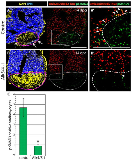
Smad3-dependent TGFβ/Activin signaling is suppressed by the SB431542 inhibitor (Alk5/4-i) after myocardial infarction. (A-B′) At 14 dpci, heart sections from transgenic fish expressing nuclear DsRed2 (red) under the cmlc2 cardiomyocyte-specific promoter were immunoassayed for Tropomyosin (TPM, blue) to visualize the intact myocardium and phospho-Smad3 (pSmad3, green). DAPI (yellow in the left panels) marks all the nuclei. The infarct is determined by the absence of Tropomyosin and DsRed2 expression (encircled by dashed lines). (A′,B′) Higher magnifications of the boxed areas in A,B. pSmad3-positive cardiomyocytes are indicated with arrowheads. (A,A′) Control regenerating hearts display numerous pSmad3-positive nuclei (green) in the injury area and in some cardiomyocytes at the infarct boundary. Arrows (A) indicate new myocardium surrounding the post-infarct. (B,B′) Treatment with 10 μM Alk5/4-i severely reduces pSmad3 staining. The injury area is larger than in the control and no invasion of cardiomyocytes is detected. (C) Quantification of pSmad3-positive cardiomyocytes in control and Alk5/4-i-treated hearts at 14 dpci. Error bars indicate s.e.m.; n=9; *P<0.01, t-test. Scale bars: 300 μm. |