|
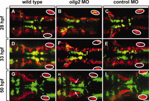
olig2 MO does not disrupt lateral line migration. All panels show dorsal views of whole embryos, anterior to the left. Elavl labeling is shown in red and Tg(isl1:GFP)rw0 reporter expression is green. A?C: 28-hpf embryos. D?F: 33-hpf embryos. G?I: 50-hpf embryos. A?F show projections of z-stack images in both red and green channels. The red channel in G?I is a single z-plane, focused on the lateral line, whereas the green channel is a z-stack image projection of the green channel. Circles mark the posterior lateral line primordia. Arrows indicate lagging facial motor neurons in olig2 MO-injected embryos. Scale bar = 30 μm.
|