Fig. 6
- ID
- ZDB-FIG-120315-34
- Publication
- Clendenon et al., 2012 - Zebrafish cadherin-11 participates in retinal differentiation and retinotectal axon projection during visual system development
- Other Figures
- All Figure Page
- Back to All Figure Page
|
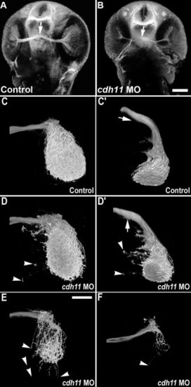
Retinotectal projections are reduced in cdh11 knockdown embryos. In 2 days postfertilization embryos, acetylated tubulin labeling detects retinal ganglion cell processes (optic nerve) and the optic chiasm (arrow). Image volumes were collected using 2-photon microscopy. A: Control embryo. B:cdh11 morphant embryo. In cdh11 knockdown embryos, retinal ganglion cell processes were present, but the optic nerve was thinner. C?F: Anterograde labeling of retinotectal projections are reduced in cdh11 knockdown embryos. Zebrafish embryos were injected with either standard control morpholino oligonucleotide (MO, C and C2) or cdh11 MO (D,D2,E,F). Embryos were fixed at 3 days postfertilization (dpf), and retinotectal projections were traced by injecting eyes with the lipophilic dye DiI (1,1-dioctadecyl-3,3,3,3-tetramethylindocarbo- cyanine perchlorate), and image volumes of the retinotectal projections were collected using 2-photon microscopy. In both controls and cdh11 knockdowns, retinal ganglion cell axons project toward the contralateral optic tectum. In control embryos (C,C2, where C2 is a rotation of the volume shown in C), upon reaching the optic tectum, retinal ganglion cells projections arborized extensively within the tectal neuropil. In slightly affected cdh11 knockdowns (D,D2), the optic nerve arborized extensively within the tectal neuropil, but there were projections that extended beyond the tectal neuropil (arrowheads). Also, there ectopic projections were detected at the optic chiasm (arrows in D and D2, where D2 is a rotation of the volume shown in D). In moderately affected embryos (E) and severely affected embryos (F), retinotectal projections were reduced, but those that reached the tectum arborized within the neuropil. In these embryos, there were also frequently projections that extended beyond the neuropil (arrowheads). Scale bars = 100 μm in A,B, 50 μm in C?F. |
| Fish: | |
|---|---|
| Knockdown Reagents: | |
| Observed In: | |
| Stage Range: | Long-pec to Protruding-mouth |

