Fig. 3
- ID
- ZDB-FIG-120315-31
- Publication
- Clendenon et al., 2012 - Zebrafish cadherin-11 participates in retinal differentiation and retinotectal axon projection during visual system development
- Other Figures
- All Figure Page
- Back to All Figure Page
|
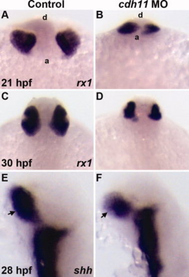
Expression of early retinal specification markers in cdh11 morphant embryos. Zebrafish embryos were injected with either standard control morpholino oligonucleotide (MO, Control) or cdh11 MO. Whole-mount in situ hybridization labeling of embryos at different stages was performed using probes for markers retinal specification. Probes are shown in the bottom right corner of each panel. A,B:rx1 labeling of 21 hours postfertilization (hpf) control and cdh11 morphant embryos. Anterior views from a ventral perspective; a, anterior embryo border; d, dorsal surface. rx1 labeling was detected in control (A) and cdh11 morphant embryos (B). C,D:rx1 labeling was strongly detected in 30 hpf control and cdh11 morphant embryos. Dorsal view of 30 hpf embryos: anterior is top. E,F: Expression of shh, a marker of retinal stalk (arrows) differentiation, was strongly detected in 28 hpf control and cdh11 morphant embryos; lateral views, anterior is up. |
| Fish: | |
|---|---|
| Knockdown Reagent: | |
| Observed In: | |
| Stage Range: | 20-25 somites to Prim-15 |

