
Fig. 2
- ID
- ZDB-FIG-110719-21
- Publication
- Muto et al., 2011 - Genetic visualization with an improved GCaMP calcium indicator reveals spatiotemporal activation of the spinal motor neurons in zebrafish
- Other Figures
- All Figure Page
- Back to All Figure Page
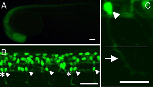
GFP expression in the SAIGFF213A;UAS:GFP double transgenic embryos. (A) A side view at 24 hpf. GFP is expressed in the central nervous system. (Scale bar, 100 μm.) (B) A side view of the spinal cord at 24 hpf observed with a confocal microscope. Arrowheads indicate CaP motor neurons. In some cases, two CaP neurons are found in one spinal segment (asterisks). (Scale bar, 100 μm.) (C) The soma (arrowhead) and axon of the GFP-expressing single CaP neuron at 24 hpf. The CaP neurons project their axons to the ventral muscle (arrow). Dotted line shows the boundary between dorsal and ventral muscles. (Scale bar, 50 μm.) |