
FIGURE
Fig. S3
- ID
- ZDB-FIG-110624-30
- Publication
- Muto et al., 2011 - Genetic visualization with an improved GCaMP calcium indicator reveals spatiotemporal activation of the spinal motor neurons in zebrafish
- Other Figures
- All Figure Page
- Back to All Figure Page
Fig. S3
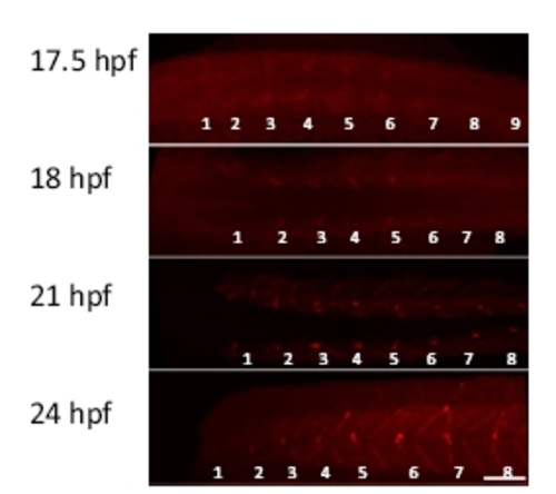
Development of neuromuscular junctions. Fluorescently labeled α-bungarotoxin was used to label neuromuscular junctions during 17.5 to 24 hours postfertilization (hpf). Dorsal views (17.5 to 21 hpf) and a lateral view (24 hpf) are shown. Myotomes are numbered from rostral to caudal (1?9). (Scale bar, 50 μm.) |
Expression Data
Expression Detail
Antibody Labeling
Phenotype Data
Phenotype Detail
Acknowledgments
This image is the copyrighted work of the attributed author or publisher, and
ZFIN has permission only to display this image to its users.
Additional permissions should be obtained from the applicable author or publisher of the image.
Full text @ Proc. Natl. Acad. Sci. USA