
FIGURE
Fig. S1
- ID
- ZDB-FIG-110624-29
- Publication
- Muto et al., 2011 - Genetic visualization with an improved GCaMP calcium indicator reveals spatiotemporal activation of the spinal motor neurons in zebrafish
- Other Figures
- All Figure Page
- Back to All Figure Page
Fig. S1
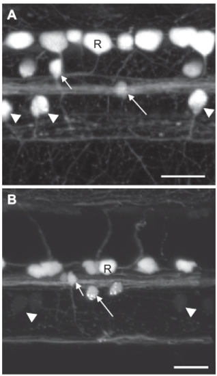
GFP expression in the SAIG213A;UAS:GFP fish at 2 days postfertilization (dpf) and 7 dpf. (A) A confocal image of the spinal neurons at 2 dpf. (B) A confocal image of the spinal neurons at 7 dpf. Note that GFP fluorescence is greatly reduced in CaP neurons. Arrowheads: CaPs; Short and long arrows: two classes of interneurons; R: Rohon-Beard neurons. (Scale bar, 20 μm.) |
Expression Data
Expression Detail
Antibody Labeling
Phenotype Data
Phenotype Detail
Acknowledgments
This image is the copyrighted work of the attributed author or publisher, and
ZFIN has permission only to display this image to its users.
Additional permissions should be obtained from the applicable author or publisher of the image.
Full text @ Proc. Natl. Acad. Sci. USA