
Fig. 1
- ID
- ZDB-FIG-110513-1
- Publication
- Li et al., 2011 - Regulation of endoderm formation and left-right asymmetry by miR-92 during early zebrafish development
- Other Figures
- All Figure Page
- Back to All Figure Page
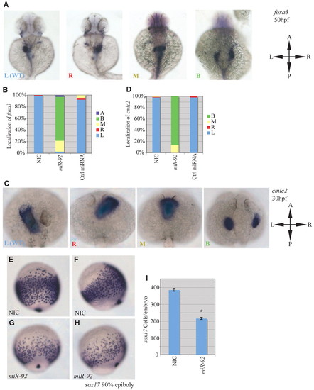
miR-92 gain of function. Gain-of-function experiments were performed by injection of single-cell zebrafish embryos with miR-92 followed by localization of specific markers, as indicated. (A) Localization of foxa3 in wild-type embryos and those injected with miR-92 at 50 hpf. Views are dorsal with anterior to the top. Images are of representative embryos with liver primordia localized to either the left (L), right (R), midline (M) or bilateral (B) positions. (B) Percentages of left, right, midline or bilateral localization of foxa3 in non-injected control (NIC) (n=188), miR-92-injected (n=37) and control miRNA-injected (n=90) embryos. In rare cases, no expression of foxa3 was detected (A, absent). (C) Localization of cmlc2 in wild type and in embryos injected with miR-92 at 30 hpf. Views are dorsal with anterior to the top. Images are of representative embryos with cardiac primordia localized to the left, right, midline or bilateral positions. (D) Percentages of left, right, midline and bilateral localization of cmcl2 in NIC (n=103), miR-92-injected (n=98) and control miRNA-injected (n=53) embryos. (E-H) Localization of sox17-expressing cells in wild-type embryos and miR-92-overexpressing embryos at 90% epiboly. (E,G) Dorsal views with anterior to the top. (F,H) Lateral views with dorsal to the right. (I) Numbers of sox17-expressing cells in NIC (n=16) and miR-92-injected embryos (n=11). Error bars represent s.e.m. *, P<0.01; Student′s t-test. A, anterior; P, posterior; L, left; R, right. |