
FIGURE
Fig. S3
- ID
- ZDB-FIG-101105-55
- Publication
- Butler et al., 2010 - Genetic and chemical modulation of spastin-dependent axon outgrowth in zebrafish embryos indicates a role for impaired microtubule dynamics in hereditary spastic paraplegia
- Other Figures
- All Figure Page
- Back to All Figure Page
Fig. S3
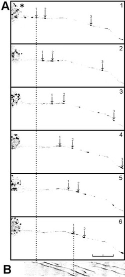
Anterograde movement of EB3-GFP puncta in a live zebrafish neuron. (A) Single frames taken from 10-minute time-lapse recording of an EB3-GFP-injected embryo (n=15) showing anterograde movement of discrete comet-like puncta of EB3-GFP fluorescence (arrows) away from the cell body (asterisk). (B) Kymograph of axon shown in A. The arrows indicate the anterograde movement of three individual comets, and the dotted lines indicate the extent of movement of one of these structures over a 10-minute period. Scale bar: 20 μm. See supplementary material Movie 1. |
Expression Data
Expression Detail
Antibody Labeling
Phenotype Data
Phenotype Detail
Acknowledgments
This image is the copyrighted work of the attributed author or publisher, and
ZFIN has permission only to display this image to its users.
Additional permissions should be obtained from the applicable author or publisher of the image.
Full text @ Dis. Model. Mech.