
FIGURE
Fig. 2
- ID
- ZDB-FIG-100616-47
- Publication
- Pang et al., 2010 - Role of G protein-coupled estrogen receptor 1, GPER, in inhibition of oocyte maturation by endogenous estrogens in zebrafish
- Other Figures
- All Figure Page
- Back to All Figure Page
Fig. 2
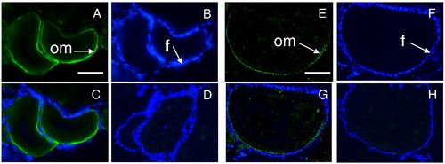
Localization of GPER in zebrafish ovarian tissue cryosection samples by immunohistochemistry using a specific GPER antibody. (A?D) Early-vitellogenic stage follicles. (E?H) Late-vitellogenic stage follicles. (A, E) Green fluorescent staining of zebrafish GPER on oocyte plasma membranes (om). (B, F) DAPI staining of nuclear DNA of follicular cells (f). (C, G) Merge. (D, H) Detection of GPER with the GPER antibody that had been neutralized with the antigen peptide and merged with DAPI stained images. Scale bar = 100 μm. |
Expression Data
| Antibody: | |
|---|---|
| Fish: | |
| Anatomical Term: | |
| Stage: | Adult |
Expression Detail
Antibody Labeling
Phenotype Data
Phenotype Detail
Acknowledgments
This image is the copyrighted work of the attributed author or publisher, and
ZFIN has permission only to display this image to its users.
Additional permissions should be obtained from the applicable author or publisher of the image.
Reprinted from Developmental Biology, 342(2), Pang, Y., and Thomas, P., Role of G protein-coupled estrogen receptor 1, GPER, in inhibition of oocyte maturation by endogenous estrogens in zebrafish, 194-206, Copyright (2010) with permission from Elsevier. Full text @ Dev. Biol.