
Fig. 6
- ID
- ZDB-FIG-100322-16
- Publication
- Kimmel et al., 2010 - Modes of developmental outgrowth and shaping of a craniofacial bone in zebrafish
- Other Figures
- All Figure Page
- Back to All Figure Page
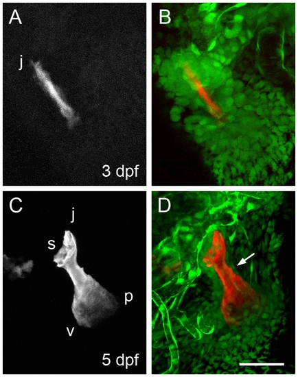
Arrangements of neural crest-derived mesenchymal cells associated with the opercle developing in the young larva. Two-color confocal imaging of live preparations. (A, C) Red channel at 3 and 5 dpf showing the Alizarin Red S labeled bone. (B, D) Merge of the red channel and the green channel showing cells expressing the fli1:eGFP transgene. Endothelial cells of capillary tubules also brightly express this transgene. The dense condensation of Op-associated cells present at 3 dpf thins out considerably by 5 dpf, particularly along the very slowing growing jp edge of the bone (arrow in D). Abbreviations and orientations as in Figure 1. Scale bar: 50 μm. |
| Gene: | |
|---|---|
| Fish: | |
| Anatomical Terms: | |
| Stage Range: | Protruding-mouth to Day 5 |