- Title
-
Modes of developmental outgrowth and shaping of a craniofacial bone in zebrafish
- Authors
- Kimmel, C.B., Delaurier, A., Ullmann, B., Dowd, J., and McFadden, M.
- Source
- Full text @ PLoS One
|
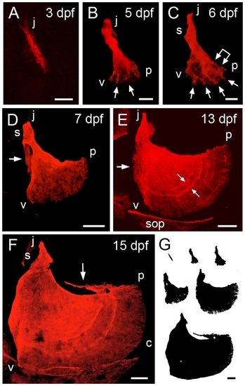
Time-course of shape changes and growth of the opercle in live, developing zebrafish larvae. A-F: Confocal projections made from z-stacks of images of live preparations vitally stained with Alizarin Red S. G: Silhouettes of the same bones scaled to the same final magnification to illustrate the amount of overall bone growth. Left-side views with dorsal approximately to the top and anterior to the left. The same orientations are used in all of the figures for this paper. (A) The Op initially ossifies as a linear bony spur. The more dorsal or j (?joint?) end is adjacent to the hyosymplectic cartilage (not shown). In occasional preparations we first see the Op as just a spot of bone at what will become the j end. (B, C) Early fan-shaped Ops at 5 and 6 dpf, with three apices j (joint apex), v (ventral) and p (posterior). We use vj, vp, and jp to describe the bone edges between these apices. The j end of the element has elaborated a joint socket (s) component of the ball-and-socket articulation the Op makes with the hyosymplectic cartilage (for anatomy see [16]). The posterior-ventral end has broadened to form a new vp edge, by developing small, secondary spurs (arrows), with intervening thinly-mineralized veils. The linked arrow in C shows the first indication of incremental bands. (D) The fan shape is expanded at 7 dpf by differential elongation of the vp edge, relative to the other two. This vp elongation continues throughout the larval period. A new veil is evident along the vj (anterior) edge (arrow). (E) The vj veil is still evident at 13 dpf (arrow). A new dorsally pointing spur has appeared at the j apex, the site of attachment of the dilator operculi muscle. Incremental bands are visible in the matrix (e.g. at the small arrow pair). The image includes portions of neighboring ossifications present at this stage, including the subopercle (sop). (F) By 15 dpf curvature of the vp edge has locally increased at one region (c). The subopercle overlaps the Op vp edge ventrally. The dorsal jp edge, where the levator operculi muscle attaches, has developed a new veil. Scale bars: 20 μm in A-C, 50 μm in D-G. |
|
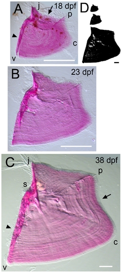
Time-course of shape changes and growth of the opercle in the late larva and in young adult fish. (A) 18 dpf. (B) 23 dpf. (C) 38 dpf. (D) Silhouettes scaled to the same final magnification to illustrate the amount of overall bone growth during this period. Imaging used oblique incident lighting to reveal matrix morphology. Incremental bands, the joint socket, and thickened, relatively heavily mineralized struts at characteristic positions along the bone are particularly well shown in C. The refractile fibers at the j apex in C are remains of the tendon that attached the dilator operculi muscle to the bone at this apex. Note a concave curvature along the vp edge (arrow in C) that was not present during the larval stages shown in Figure 1. Abbreviations and orientations as in Figure 1. Scale bars: 200 μm. |
|
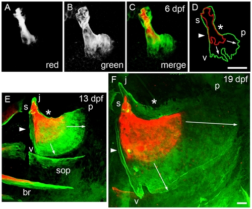
Mineralized matrix addition occurs in a stereotyped spatiotemporal pattern. Live confocal imaging as in Figure 1. The larvae were vitally stained successively with two Ca2+-binding dyes, first Alizarin Red S, then Calcein (green), with a washout period between the two applications. They were imaged just after the second staining period. (A-D) The same Op, stained first for 2 hr at 3 dpf (A, red channel), and then overnight between 5 and 6 dpf (B, green channel). C shows the merge and D the outlines of the two colors. The vj edge (arrowhead in D) and jp edge (asterisk) show only a thin layer of new (green) matrix on top of the older (red) matrix. In contrast, the vp edge, showing short spurs at the time of the first pulse, grows out prominently (arrows in D). (E) Another preparation in which a larva was stained with overnight first at 6?7 dpf and then at 12?13 dpf. The jp edge, as at the earlier stage, shows only a very thin layer of new (green) bone (asterisk). The veil along the vj edge (arrowhead), and the upward pointing short j apex spur are made of new bone. Note that this two-color matrix staining method also reveals that mineralization of the branchiostegal ray (br) began before 7 dpf (since the br is doubly labeled), but that subopercle (sop) mineralization is initiated only after day 7 (since the sop is not Alizarin Red-labeled). (F) A preparation stained overnight first at 12?13 dpf and then at 18?19 dpf. In contrast to the earlier stages, there is now an elaborate outgrowing veil, made of new bone, along dorsal jp edge. The anterior strut along the vj edge is new (arrowhead), and the j apex has elongated by new bone addition. Note that in both E and F the outgrowth of the vp edge is differential, more rapid near the p apex than near the v apex, as indicated by the lengths of the arrows. Abbreviations and orientations as in Figure 1. Scale bars: 40 μm. |
|
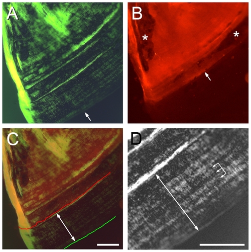
The Op of the young adult shows a high rate of incremental banding outgrowth of the vp edge. (A) Image at 54 dpf with green monochromatic (to increase resolution) transmitted light, and crossed-polarizing filters to reveal birefringence and the incremental banding pattern. The arrow indicates the vp edge of the bone. (B) Epiflorescence of Alizarin Red S, applied as a pulse at 43 dpf, in the same field as in A. The labeling front (arrow) shows where the vp edge was located at the 43 dpf stage. Sites (Howship′s lacunae) of likely remodeling (bone resorption by osteoclasts, followed by replacement with new, unlabeled bone) in the old bone behind this front are indicated by asterisks. (C) Merge of A and B. The Alizarin Red front is indicated by the red line, the bone vp edge by the green line, and the double-headed arrow shows the approximate extent of outgrowth during the 11 day interval after labeling (an average 333 Ám, from several measurements along the bone). (D) Detail of the banding pattern between the labeling front and the bone edge (double-headed arrow). Widths of prominent bands are about 30 μm; the three linked arrows show two 30 μm intervals. We note more finely spaced bands are also present, and that we measured a substantial variation in bandwidths among different preparations. Scale bars: 200 μm. |
|
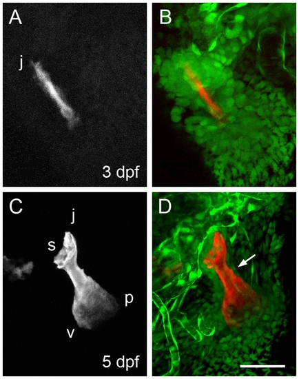
Arrangements of neural crest-derived mesenchymal cells associated with the opercle developing in the young larva. Two-color confocal imaging of live preparations. (A, C) Red channel at 3 and 5 dpf showing the Alizarin Red S labeled bone. (B, D) Merge of the red channel and the green channel showing cells expressing the fli1:eGFP transgene. Endothelial cells of capillary tubules also brightly express this transgene. The dense condensation of Op-associated cells present at 3 dpf thins out considerably by 5 dpf, particularly along the very slowing growing jp edge of the bone (arrow in D). Abbreviations and orientations as in Figure 1. Scale bar: 50 μm. EXPRESSION / LABELING:
|
|
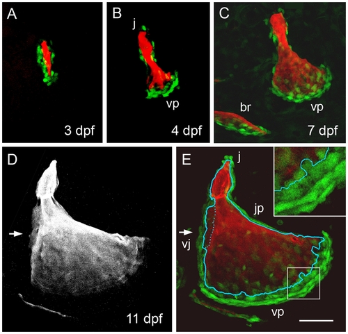
Osteoblast arrangements change dynamically during opercle morphogenesis. Confocal imaging of live preparations. (A-C) Two-color merged images showing Alizarin Red A labeling of the matrix and osx:eGFP labeling of the bone forming cells. (A) At 3 dpf osteoblasts line up along the developing bony spur. (B, C) At 4 and 7 dpf a new arrangement is present with cells especially concentrated along the rapidly outgrowing vp edge. The newly forming posterior branchiostegal ray (br) is included in C. (D) Red channel, and (E) merge at 11 dpf. Arrows indicate the vj veil. In E the outline of the Alizarin Red labeled bone (from D) is superimposed. Very flattened and compact looking osx:eGFP-expressing cells line the very slowly growing jp edge and are present in outer rows of the very rapidly growing vp edge. Small round cells are present at the j apex spur. Larger diffusely labeled cells are present along the vj veil and in the innermost row along the vp edge, where the cells immediately contact new mineralized matrix (a portion of this edge is enlarged in the inset). Scale bar: 50 μm. EXPRESSION / LABELING:
|
|
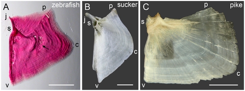
Comparison of opercle shapes of (A) zebrafish, Danio rerio, (B) sucker, Castostomus sp., and (C) northern pike, Esox lucius. Letters along the edges indicate hypothetically homologous locations along the bones. Shape differences along the upper (jp) edge and the j apex are discussed in the text. The arrow in (A) indicates a prominent Howship′s lacunae, a site of osteoclast-mediated bone resorption. The vp edge appears to outgrow differentially in all three species, as indicated by the incremental bands being wider in region c. The region between c and p shows the narrowest banding for both zebrafish and sucker, resulting in the concavity here shared by these two species but not by the pike. Zebrafish and the sucker are placed in separate families within the order Cypriniformes, the pike is in the order Esociformes and therefore is an outgroup to the other two species. Scale bars: 0.5 mm (A), 10 mm (B,C). |

