
Fig. S3
- ID
- ZDB-FIG-100309-38
- Publication
- Gutzman et al., 2010 - Epithelial relaxation mediated by the myosin phosphatase regulator Mypt1 is required for brain ventricle lumen expansion and hindbrain morphogenesis
- Other Figures
- All Figure Page
- Back to All Figure Page
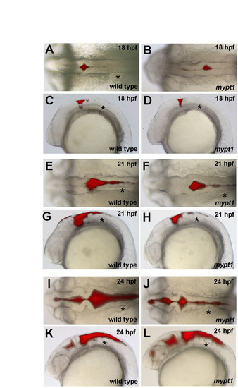
Ventricle injection time course of wild type and mypt1 mutants. Gross morphology was analyzed by ventricle injection followed by brightfield and fluorescence microscopy (n>10 for each panel). (A-D) At 18 hpf, wild-type and mypt1 mutant embryos had similar brain morphology. (A,B) Both had an initial ventricle opening at the r0/r1 boundary. (C,D) Rhombomere morphology was apparent in the lateral view. (E-H) At 21 hpf, hindbrain formation in mypt1 mutants also looked similar to that in wild-type embryos. (E,F) The hindbrain was separated within r1 and r2, and open in more posterior regions but not fully separated. (G,H) At this time, the rhombomeres were also prominent as seen from the lateral view and appeared normal in mypt1 mutants. (I-L) At 24 hpf, a clear defect was apparent in mypt1 mutants compared with the wild type, both dorsally (I,J) and laterally (K,L). Asterisk indicates ear. |