|
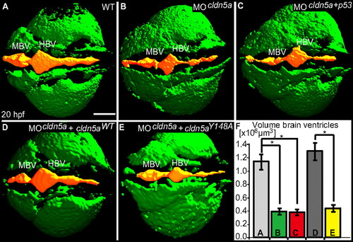
Loss of Claudin5a affects expansion of the brain ventricular lumen at 20 hpf. (A–E) Shown are confocal microscopic Z-scans of in vivo sodium green indicator-labeled embryos. The ventricular lumen is indicated by false-coloring (orange). Indicated are ventricles of the midbrain (MBV) and hindbrain (HBV). (F) Quantifications of ventricular volume were generated from detailed 3D-reconstructions of confocal Z-scan sections for a total of 4–5 embryos per sample group by using Volocity software (unit size is [x106 μm3]). Data represent mean ± SEM, n ≥ 4, *, P = 0.016 in all three cases. (Scale bar: 100 μm.)
|