
Fig. S4
- ID
- ZDB-FIG-100302-48
- Publication
- Zhang et al., 2010 - Establishment of a neuroepithelial barrier by Claudin5a is essential for zebrafish brain ventricular lumen expansion
- Other Figures
- All Figure Page
- Back to All Figure Page
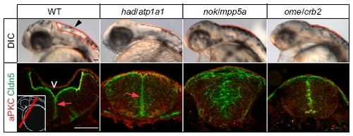
Phenotypic comparison of brain ventricle expansion defects in different zebrafish ion pump Atp1a1-deficient or cell polarity mutants. Arrowhead indicates location of the hindbrain ventricle in a WT embryo at 30 hpf. Hindbrain ventricular shapes are outlined in red. Sections of immunohistochemical stainings of the brain neuroepithelium show that Cldn5a colocalizes apically together with atypical protein kinase C (aPKC) at the inner ventricular (V) surface at 30 hpf (arrows; Left Inset indicates the cross-sectional plane through the hindbrain region). Localization of Cldn5a and aPKC is affected in nokm520/mpp5a and omem289/crb2 cell polarity mutants. (Scale bar: 50 μm.) |