Fig. 1
- ID
- ZDB-FIG-100302-11
- Publication
- Jing et al., 2010 - Temporal and spatial requirements of unplugged/MuSK function during zebrafish neuromuscular development
- Other Figures
- All Figure Page
- Back to All Figure Page
|
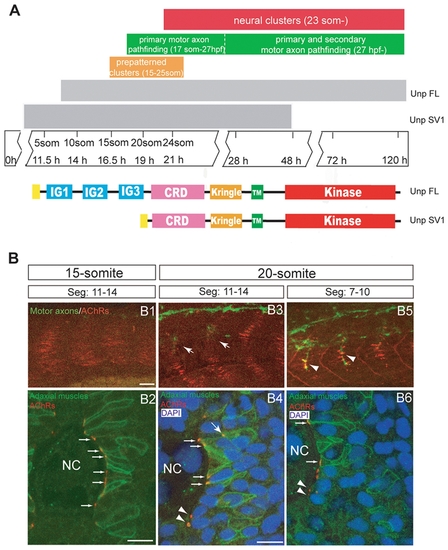
Neuromuscular synaptogenesis in zebrafish. (A) Schematic representation of the different steps during neuromuscular synapse development. unplugged SV1 mRNA is expressed transiently from the tailbud stage to 48 hpf, while unplugged FL mRNA is expressed from the 10-somite stage and continues to be expressed in adult muscle. Bottom: Domain structure of the unplugged FL and SV1 isoforms. (B) Synaptogenesis at the 15- or 20-somite stage. 15-somite (B1) or 20-somite stage (B3 and B4) embryos stained for motor axons (znp-1, green) and AChR clusters (α-BTX, red). In segments 11?14, AChR prepatterned clusters appear at the 15-somite stage before motor neurons have exited the spinal cord (B1); prepatterned clusters coalesce and motor axons (arrows) begin to approach the myotome at the 20-somite stage (B3). In anterior older segments 7?10, motor axons have contacted non-migratory adaxial cells (arrowheads) at the 20-somite stage (B5). (B2, B4, B6) Cross-sectional views of Tg(smyhc1:mcherry-CAAX) embryos, which express mCherry specifically on the membrane of adaxial cells, stained for adaxial muscles (anti-DsRed, green), prepatterned clusters (α-BTX, red) and nuclei (DAPI, blue). The same segments as in (B1, B3, B5) were analyzed. (B2) At the 15-somite stage, adaxial muscles remain adjacent to notochord, and prepatterned clusters are localized at the medial side of the cells (small arrows). (B4) At the 20-somite stage, adaxial muscles have migrated laterally. Some prepatterned clusters co-migrate with the adaxial cells (big arrow). Prepatterned clusters appear on the medial side of fast muscle fibers (arrow heads). Prepatterned clusters remain on the medial side of the non-migratory adaxial cells (small arrows). (B6) In older segments 7?10, adaxial muscles have migrated further away; AChR clusters remain on the medial side of non-migratory adaxial cells (small arrows). AChR clusters appear on the medial side of fast muscle fibers (arrow heads). NC: notochord. Scale bars: 20 μM in B; 10 μM in B2 and B4. |

