
Fig. 5
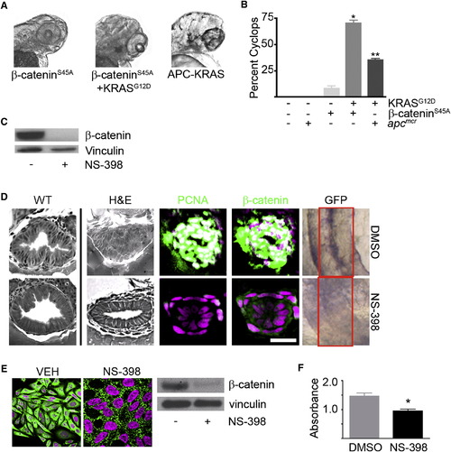
KRAS-Mediated Intestinal Cell Proliferation following Loss of APC Requires β-Catenin (A) Zebrafish embryos were injected with β-cateninS45A mRNA (left) along with KRAS mRNA (middle). Also shown is a representative image of the APC-KRAS embryo (right). At 72 hpf, the embryos were fixed and photographed. (B) The percent cyclops was analyzed (*p < 0.01 WT versus β-catenin-KRAS, **p < 0.01 WT versus APC-KRAS). (C) Protein was harvested from apcmcr embryos treated with DMSO or NS-398 and subjected to western blot analysis for β-catenin (top) or vinculin (bottom). (D) Wild-type uninjected or apcmcr embryos injected with KRAS mRNA treated with VEH (top) or NS-398 (bottom) were stained by H&E (WT, left; APC-KRAS, right) and for DNA (magenta), PCNA (green), and β-catenin (green). TOPGFP-APCmo-KRAS embryos were stained for GFP expression (purple). Boxes indicate the intestine. (E) SW-480 cells treated with DMSO or NS-398 were stained for DNA (magenta) and β-catenin (green). Protein lysates were subjected to western blot analysis for β-catenin (top) and vinculin (bottom). (F) Cells were subjected to MTT analysis (*p < 0.05 versus DMSO; error bars, SEM). Overlapping expression is shown in white. All images were captured using the same exposure and represent at least three independent experiments. Scale bar, 10 μm. |
Reprinted from Cell, 137(4), Phelps, R.A., Chidester, S., Dehghanizadeh, S., Phelps, J., Sandoval, I.T., Rai, K., Broadbent, T., Sarkar, S., Burt, R.W., and Jones, D.A., A two-step model for colon adenoma initiation and progression caused by APC loss, 623-634, Copyright (2009) with permission from Elsevier. Full text @ Cell