
Fig. 3
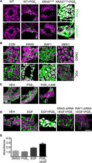
KRAS and RAF1 Direct Stabilized β-Catenin to the Nucleus (A) WT and KRAS-injected zebrafish embryos treated with PGE2 were stained for DNA (magenta), PCNA (green), and β-catenin (green). (B) Human 293 cells were transfected with constitutively active KRAS, RAF1, or MEK1 and either DMSO (top) or PGE2 (bottom). Cells were stained for DNA (magenta) and β-catenin (green). (C) 293 cells treated with DMSO, PGE2 or PGE2, and leptomycin B were stained for DNA (magenta) and β-catenin (green). (D) 293 cells were transfected with control or KRAS- or RAF1-directed siRNA and treated with EGF and PGE2 or vehicle and then stained for DNA (magenta) and β-catenin (green). (E) 293 cells were subjected to an MTT assay (*p < 0.05 versus DMSO; error bars, SEM). Overlapping expression is shown in white. All images were captured using the same exposure and represent three independent experiments. Scale bar, 10 μm. |
Reprinted from Cell, 137(4), Phelps, R.A., Chidester, S., Dehghanizadeh, S., Phelps, J., Sandoval, I.T., Rai, K., Broadbent, T., Sarkar, S., Burt, R.W., and Jones, D.A., A two-step model for colon adenoma initiation and progression caused by APC loss, 623-634, Copyright (2009) with permission from Elsevier. Full text @ Cell