
Fig. 3
- ID
- ZDB-FIG-090224-48
- Publication
- Dowling et al., 2009 - Loss of myotubularin function results in T-tubule disorganization in zebrafish and human myotubular myopathy
- Other Figures
- All Figure Page
- Back to All Figure Page
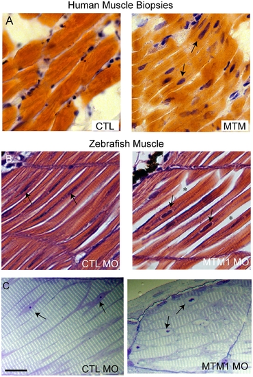
Abnormal histopathology in 72 hpf myotubularin morphants. (A) H/E stained longitudinal myofibers from myotubular myopathy (MTM) and age matched control (CTL) human muscle biopsies. Arrows point to abnormal nuclei. (B) H/E stained longitudinal myofibers from control (CTL MO) and myotubularin (MTM MO) morphant 72 hpf embryos. Myonuclei are abnormally rounded (arrows), and there is increased space between fibers (*). (C) Toluidine blue stained semi-thin sections from 72 hpf morphants. Myonuclei from myotubularin morphants are large, abnormally rounded, and contain discrete nucleoli (arrows). Sarcomeric units, however, are normal in appearance. Scale bar = 20 mm. |
| Fish: | |
|---|---|
| Knockdown Reagent: | |
| Observed In: | |
| Stage: | Protruding-mouth |