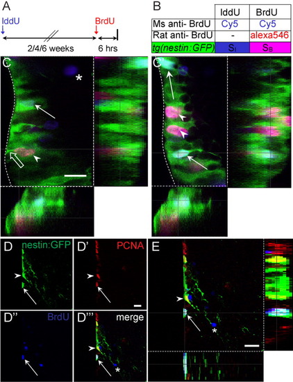
Tg[-3.9nestin:GFP] is expressed in cycling and long-term label retaining cells at the ventricular zone. A,B: Summary of experimental strategy to detect proliferating cells. C,C′: Two confocal z-planes of a single transverse section through the ventral telencephalon of tg[-3.9nestin:GFP]. Iododeoxyuridine (IddU) -labeled cells were left to survive for 4 weeks followed by a pulse of bromodeoxyuridine (BrdU) for 6 hr before killing. Arrowheads indicate colocalization of green fluorescent protein (GFP, green) with fast-cycling BrdU-labeled cells (pink), whereas open arrow shows the endfoot at the ventricular zone (VZ). Arrows and asterisks indicate IddU-labeled cells (blue) that are either GFP-positive or -negative, respectively. Dotted line delineates boundary of the VZ. D-D′″: Confocal images of single and merged channels of tg[-3.9nestin:GFP] positive cells in the ventral telencephalon. There is colocalization of GFP+/proliferating cell nuclear antigen-positive (PCNA+) cells (arrowheads) and GFP+/PCNA+/BrdU+ cells after a 2-week chase period (arrows), indicating that tg[-3.9nestin:GFP] is expressed in cycling cells and progenitors that are capable of self-renewal. Some BrdU+/PCNA- cells have migrated out of the VZ (asterisks) and are likely to be differentiated. E: Single optical z-section clearly reveals a triple stained GFP+/PCNA+/BrdU+ self-renewing progenitor. Scale bar 10 μm in C, 25 μm in D′, E.
|
