|
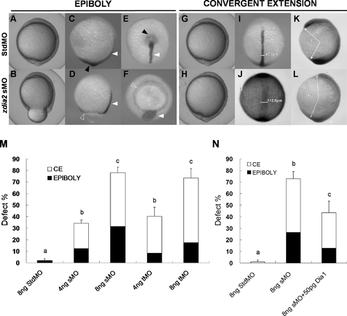
Knockdown of zdia2 by MO interferes with gastrulation cell movements in a dose-dependent manner. Embryos injected with 8 ng of standard control (std) MO (the upper row) and zdia2 sMO (the bottom row) were examined and photographed under a stereomicroscope at 10 hpf. Embryos were then fixed, stained by a no tail riboprobe (C, D, K and L, side view; E and F, vegetal view; I and J, dorsal view) or in combination with a goosecoid riboprobe and photographed (K and L, side view). Tail buds and notochords are respectively indicated by black and white arrowheads (C?F). The zdia2 sMO caused incomplete epiboly formation with a ring structure near the vegetal pole as indicated by the ntl staining (D and F, open arrowheads). Widths of the notochords were measured and labeled in I and J. Lengths of the body axis were also estimated by the angle (labeled) between the prechordal plate and tail bud as described in the text (K and L). (M) Different MOs at 4 or 8 ng as designated were injected, and the embryos were examined at 10 hpf. Each treatment was repeated at least three times and analyzed as described, and only upper error bars of standard deviations are shown. (N) Embryos were injected with stdMO, zdia2 sMO or zdia2 sMO with 50 pg mdia mRNA as indicated. Epiboly and convergent extension defects were determined, analyzed, and presented as described in (M). Different letters on top of each column indicate a significant difference between treatments (P<0.05).
|