|
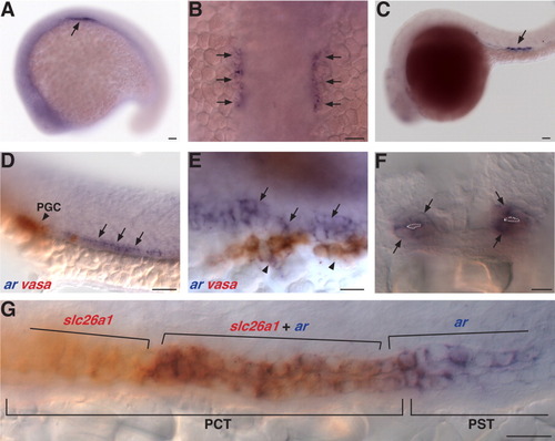
Expression of the ar gene in the embryonic pronephros. A,B: ar transcripts are detected in a bilateral row of cells (arrows) lateral to the somites at the 18 somite stage. C: ar expression is displaced posteriorly (arrow) by 24 hours (h). D,E: Double-label in situ hybridization of ar (blue, arrows) and vasa (red, arrow heads), a marker of primordial germ cells, at 24 h. E: Close association between vasa and ar expressing cells. F: Coronal section showing ar expression (arrows) in cells that form pronephric tubules at 24 h. Tubule lumen outlined in white, dorsal to the top. G: A subset of cells in the proximal convoluted tubule (PCT) express both ar (blue) and slc26a1 (red), a gene expressed in the PCT but not in the adjacent proximal straight tubule (PST), at 24 h. ar expression is also present in the presumptive PST. A,C-E,G, lateral views anterior to the left, B, dorsal view, anterior to the top. Scale bars = 50 μm in A-D, 10 μm in F, 20 μm in E,G.
|

