Fig. 3
|
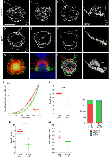
Two distinct axon arbor morphologies formed by habenular projection neurons. (a-g, i) Dorsal, (c′, f′, i′) lateral and (h) transverse confocal images of the IPN region in 4 dpf larvae. In all panels habenular axons were labeled by electroporation except for (h) where lipophilic dye tracing was used to visualize axon terminals. Panels show three-dimensional reconstructions except (h), which is a single-depth confocal image. (a-c′) Three examples of L-typical (L-typ) arbors. These arbors are shaped like a domed crown and arborize over a considerable DV extent (compare dorsal (c) and lateral (c′) views of an example L-typical arbor). See also Additional file 2. (d-f′) Examples of R-typical (R-typ) arbors, which are considerably flatter. See also Additional file 3. Panel (d) shows two R-typical arbors formed by two R habenular neurons. In (c′, f′) orange dotted lines and bars indicate how DV extent was measured for L-typical and R-typical arbors (see Materials and methods). (g) Schematic to illustrate how the radial distribution of axon density was measured. The perimeter of the arbor (indicated by white line) was defined using the convex hull method. The area covered by the arbor was then divided into ten equally spaced concentric shells (colored white-red) centered on the centroid of the hull. After thresholding, the number of pixels representing axon branches was then quantified for each shell (see Materials and methods). (h) Transverse confocal section through the IPN showing the entire contingent of L and R habenular axon terminals labeled using DiD (red) and DiI (green), respectively, in a Tg(h2afz-GFP) transgenic larva to label the nuclei of IPN neurons (blue). Most L-sided neurons innervate the dIPN whereas the majority of R-sided axons terminate in the vIPN. (i, i′) L- and R-sided axons labeled in the same larva. L-typical arbors are located dorsal to R-typical arbors. (j) Cumulative fraction of axon density plotted against radius (measured from centroid of the convex hull to the perimeter) for ten L-typical and seven R-typical arbors. The data are fit by fourth-order polynomial models (solid lines). Dotted lines indicate the 95% confidence band of each best-fit curve. R-typical arbors have a greater proportion of axon density localized towards the perimeter of the arbor. (k) L-typical arbors extend over a greater DV extent than R-typical arbors. (l) L-typical arbors have more branch points than R-typical arbors. (m) The width/length ratio of L-typical arbors shows a trend towards higher values than for R-typical arbors. (n) The majority of L-sided habenular neurons elaborate L-typical arbors whereas most R-sided neurons form R-typical axon arbors. Horizontal lines indicate mean values and error bars show standard error of the mean. ***p < 0.001. |

