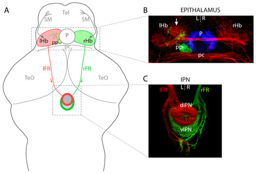
Left-right asymmetries in the telencephalon-habenula-IPN pathway. (a) Schematic of a 4 dpf larval zebrafish brain. The bilaterally paired epithalamic habenular (Hb) nuclei receive afferent inputs from nuclei in the telencephalon (Tel) via the stria medullaris (SM; grey arrows; asymmetric innervation of the R habenula [20] is not indicated) and diencephalon (not shown). Habenular neurons send efferent projections via the fasciculi retroflexus (FR) to the interpeduncular nucleus (IPN) in the ventral midbrain. L- and R-sided habenular axons are segregated along the DV axis of the IPN wherein the L habenula predominantly innervates the dIPN and makes less substantial projections to the vIPN whereas the vast majority of R-sided axons terminate in the vIPN. In addition to the habenulae, the epithalamus contains the pineal complex, comprising the photoreceptive pineal (P) and parapineal (pp) nuclei. The parapineal is located on the L side of the dorsal midline and projects efferent axons that exclusively innervate the L habenula. (b) Neuroanatomical asymmetries in the epithalamus. Anti-acetylated tubulin immunostaining (red) reveals the L habenula displays a greater density of neuropil, especially in the dorso-medial aspect of the nucleus (arrow). The pineal (blue) and parapineal (green) nuclei are visualized by expression of GFP in a Tg(foxD3:GFP) transgenic larva. Parapineal efferent axons predominantly terminate in the asymmetric medial neuropil of the L habenula. (c) Three-dimensional reconstruction of habenular axon terminals in the ventral midbrain, labeled using lipophilic carbocyanine dyes applied to the habenulae. L-sided axons were labeled with DiD (red) and R-sided axons with DiI (green). The dIPN is almost exclusively innervated by L-sided axons, whereas the ventral target receives a majority of R-sided inputs. All panels show dorsal views, anterior top. pc, posterior commissure; TeO, optic tectum.
|

