
Fig. 4
- ID
- ZDB-FIG-080508-36
- Publication
- Henry et al., 2001 - Roles for zebrafish focal adhesion kinase in notochord and somite morphogenesis
- Other Figures
- All Figure Page
- Back to All Figure Page
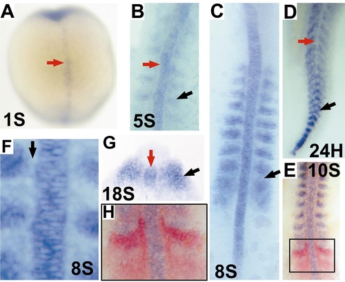
fak mRNA is expressed in the notochord, somite, and unsegmented paraxial mesoderm in wild-type zebrafish embryos. (A?F, H) Dorsal view. (G) Transverse view. ?S? indicates somite stages. (A, B) Expression of fak in the notochord (red arrows) and somites (black arrows) in early embryos. (C) The black arrow points to the posterior border of the eighth (last formed) somite. (D) Expression of fak in somites (black arrow) in a late embryo. The absence of notochordal fak is indicated at the red arrow. (E, H) Two-color in situ hybridization (H is enlarged view of the box region in E) of the localization of fak (blue) and the two strong bands of papc expression (red) in the unsegmented paraxial mesoderm. (F) fak is not expressed in the adaxial cells that abut the notochord (black arrow). (G) A cross-section of an 18-somite embryo indicates that fak is expressed throughout the region of the notochord that is still undergoing cell intercalation (red arrow) and throughout the dorsal?ventral extent of the somites (black arrow). |
| Gene: | |
|---|---|
| Fish: | |
| Anatomical Terms: | |
| Stage Range: | 1-4 somites to Prim-5 |
Reprinted from Developmental Biology, 240(2), Henry, C.A., Crawford, B.D., Yan, Y.-L., Postlethwait, J., Cooper, M.S., and Hille, M.B., Roles for zebrafish focal adhesion kinase in notochord and somite morphogenesis, 474-487, Copyright (2001) with permission from Elsevier. Full text @ Dev. Biol.