Fig. 5
- ID
- ZDB-FIG-080508-37
- Publication
- Henry et al., 2001 - Roles for zebrafish focal adhesion kinase in notochord and somite morphogenesis
- Other Figures
- All Figure Page
- Back to All Figure Page
|
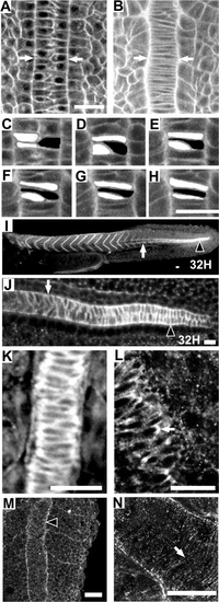
Zebrafish notochord cells intercalate as in Xenopus embryos; Fak protein is highly expressed in the region of the notochord that is undergoing convergence; and Fak is phosphorylated at the notochord-somite boundary. Anterior is towards the top or left in all panels. Dorsal views except (I) and (J), which are side views. (A–H) Time-lapse analysis of a vitally-stained zebrafish embryo (see methods) from the 1-somite stage through the 4-somite stage at the level of the first 4 somites. At time 0 min (A), the notochord is two cells wide (white arrows) and by time 38 min (B) cellular intercalation results in a notochord that is one cell wide. (C–H) Enlarged view of three cells intercalating. (C) 0 min. (D) 10 min. (E) 23 min. (F) 30 min. (G) 35 min. (H) 40 min. (I–L) Immunostaining for Fak protein. The posterior region of the notochord undergoing cell intercalation is noted with an arrowhead. White arrow indicates the notochord–somite boundary anterior to the intercalating domain. (K) A 10-somite embryo, dorsal view at the level of somite 4 showing the notochord–somite boundary. (L) Enlarged view of a notochord undergoing intercalation in a 10-somite embryo, at the level of somite 8. An arrow notes a discrete plaque of Fak. (M, N) Immunostaining for pY397FAK. M: A 13-somite embryo at somite 13 stained for phospho-Fak at the notochord-somite boundary (black arrowhead). (N) Projection of confocal sections that include the entire thickness of notochord of a 13-somite embryo showing circumferential striations in phospho-Fak staining perpendicular to the notochord axis. Scale bars, 20 μm, with the exception of (I), where scale bar is 40 μm. |
Reprinted from Developmental Biology, 240(2), Henry, C.A., Crawford, B.D., Yan, Y.-L., Postlethwait, J., Cooper, M.S., and Hille, M.B., Roles for zebrafish focal adhesion kinase in notochord and somite morphogenesis, 474-487, Copyright (2001) with permission from Elsevier. Full text @ Dev. Biol.

