
FIGURE
Fig. S2
- ID
- ZDB-FIG-080502-31
- Publication
- Farooq et al., 2008 - Histone deacetylase 3 (hdac3) is specifically required for liver development in zebrafish
- Other Figures
- All Figure Page
- Back to All Figure Page
Fig. S2
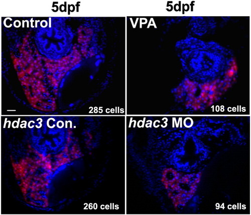
Reduction of liver cells in VPA-treated embryos and hdac3 morphants. Embryos from Tg(lfabp:RFP; elaA:EGFP) are cross-sectioned and liver cells of the largest size section (middle of the liver) are counted under a microscope after DAPI staining. RFP positive and DAPI positive cells are counted as liver cells. Number of cells is indicated at the right bottom corner of each panel and it is the average from 3 embryos of the sample group. Scale bar is 50 μm. |
Expression Data
| Gene: | |
|---|---|
| Fish: | |
| Condition: | |
| Knockdown Reagent: | |
| Anatomical Term: | |
| Stage: | Day 5 |
Expression Detail
Antibody Labeling
Phenotype Data
Phenotype Detail
Acknowledgments
This image is the copyrighted work of the attributed author or publisher, and
ZFIN has permission only to display this image to its users.
Additional permissions should be obtained from the applicable author or publisher of the image.
Reprinted from Developmental Biology, 317(1), Farooq, M., Sulochana, K.N., Pan, X., To, J., Sheng, D., Gong, Z., and Ge, R., Histone deacetylase 3 (hdac3) is specifically required for liver development in zebrafish, 336-353, Copyright (2008) with permission from Elsevier. Full text @ Dev. Biol.