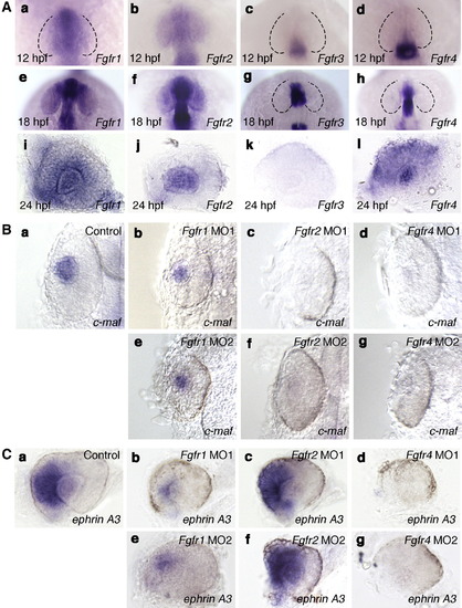
Expression pattern and function of Fgfrs in the eye. A: The expression patterns of Fgfr1 (a, e, i), Fgfr2 (b, f, j), Fgfr3 (c, g, k) and Fgfr4 (d, h, l) at 12 hpf (a?d), 18 hpf (e?h) and 24 hpf (i?l). (a, e) The expression of Fgfr1 was detected in the future nasal retina at 12 and 18 hpf. (i) At 24 hpf, the expression of Fgfr1 was detected in the lens and inner retina. (b) Fgfr2 expression was detected in the entire optic vesicle at 12 hpf. (f) At 18 hpf, Fgfr2 expression was detected in the lens. (j) By 24 hpf, Fgfr2 expression was detected at high levels in the lens. (c, g, k) The expression of Fgfr3 was not detected in the eye at 12, 18 and 24 hpf. (d, h) Fgfr4 expression was not detected in the eye at 12 and 18 hpf. (l) The expression of Fgfr4 was detected in the retina and lens at 24 hpf. (a?h) Dorsal views with anterior to the top. (i?l) Lateral views with anterior to the left and dorsal to the top. B: Expression of c-maf in wild-type embryos (a) and embryos injected with Fgfr1 MO1 (b) or MO2 (e), Fgfr2 MO1 (c) or MO2 (f) and Fgfr4 MO1 (d) or MO2 (g) at 24 hpf. The expression pattern of c-maf in the embryos injected with Fgfr1 MO1, Fgfr2 MO1 or Fgfr4 MO1 was essentially similar to that in embryos injected with Fgfr1 MO2, Fgfr2 MO2 or Fgfr4 MO2, respectively. (b, e) The expression of c-maf was reduced slightly in the lens region of Fgfr1 MO1- or MO2-injected embryos compared with wild-type embryos. (c, d, f, g) In the lens region of Fgfr2 MO1- or MO2-injected embryos or Fgfr4 MO1- or MO2-injected embryos, the expression of c-maf was not detected. Dorsal views with anterior to the top. C: Expression of ephrin A3 in wild-type embryos (a) and embryos injected with Fgfr1 MO1 (b) or MO2 (e), Fgfr2 MO1 (c) or MO2 (f) and Fgfr4 MO1 (d) or MO2 (g) at 28 hpf. The expression pattern of ephrin A3 in embryos injected with Fgfr1 MO1, Fgfr2 MO1 or Fgfr4 MO1 was essentially similar to that in embryos injected with Fgfr1 MO2, Fgfr2 MO2 and Fgfr4 MO2, respectively. (b, e) The expression of ephrin A3 was reduced in the nasal retina of Fgfr1 MO1- or MO2-injected embryos compared with control embryos. (c, f) In Fgfr2 MO1- or MO2-injected embryos, ephrin A3 expression was unaffected. (d, g) The expression of ephrin A3 was not detected in Fgfr4 MO1- or MO2-injected embryos. Lateral views with anterior to the left and dorsal to the top.
|

