|
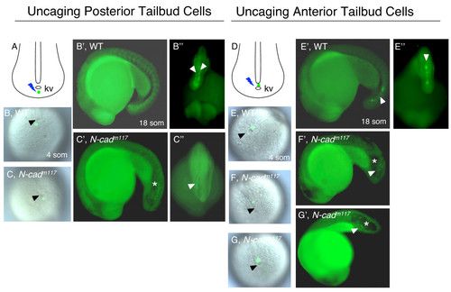
Impaired movement of anterior tailbud cells in N-cadm117 mutants. Posterior (A-C″) and anterior (D-G&prime) cells in the tailbud were uncaged at 4 som (A-C,D-G) and imaged at 18 som (B′-C″,E′-G′). (A,D) Schematic diagrams of a dorsal view of the tailbud at 4 som, illustrating where the uncaging was done (green dot). (B,C,E-G) Dorsal views of the tailbud at 4 som, showing where the uncaging was done (green label), in WT (B,E) and N-cadm117 mutants (C,F,G). Lateral (B′,E′) and dorsal (B″,E″) views of 18som WT embryos indicating the position of uncaged cells (white arrowheads). Lateral (C′,F′,G′) and dorsal (C″) views of N-cadm117 mutants indicating position of labeled cells (white arrowheads). Abbreviations and symbols: som, somite; kv, Kupffer′s vesicle; asterisk indicates the position of the vacuole in the tailbud, black arrowheads show KV.
|