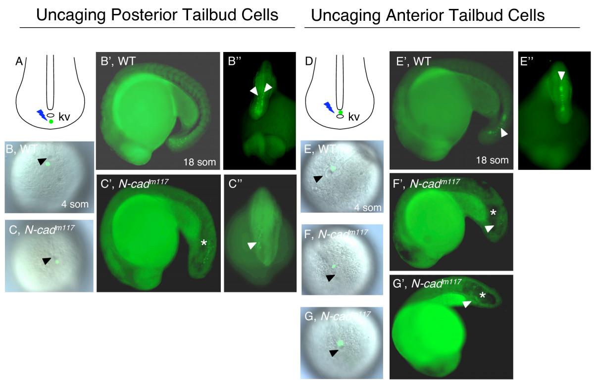
Fig. 7 Impaired movement of anterior tailbud cells in N-cadm117 mutants. Posterior (A-C″) and anterior (D-G&prime) cells in the tailbud were uncaged at 4 som (A-C,D-G) and imaged at 18 som (B′-C″,E′-G′). (A,D) Schematic diagrams of a dorsal view of the tailbud at 4 som, illustrating where the uncaging was done (green dot). (B,C,E-G) Dorsal views of the tailbud at 4 som, showing where the uncaging was done (green label), in WT (B,E) and N-cadm117 mutants (C,F,G). Lateral (B′,E′) and dorsal (B″,E″) views of 18som WT embryos indicating the position of uncaged cells (white arrowheads). Lateral (C′,F′,G′) and dorsal (C″) views of N-cadm117 mutants indicating position of labeled cells (white arrowheads). Abbreviations and symbols: som, somite; kv, Kupffer′s vesicle; asterisk indicates the position of the vacuole in the tailbud, black arrowheads show KV.
Image
Figure Caption
Figure Data
Acknowledgments
This image is the copyrighted work of the attributed author or publisher, and
ZFIN has permission only to display this image to its users.
Additional permissions should be obtained from the applicable author or publisher of the image.
Full text @ BMC Dev. Biol.

