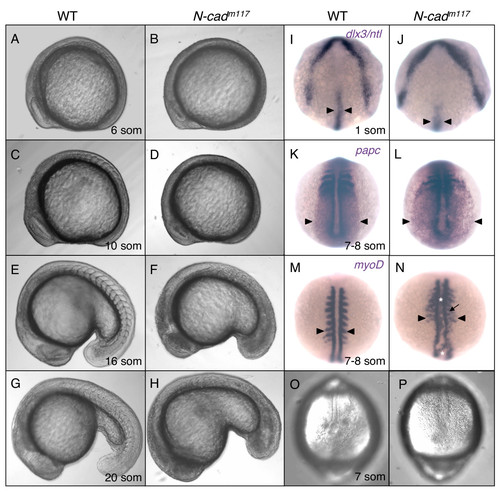
Mesodermal morphogenesis defects in N-cadm117 mutants. (A-H) Lateral views of WT (A,C,E,G) and N-cadm117 mutant (B,D,F,H) embryos imaged with Nomarski optics at 6 som (A,B), 10 som (C,D), 16 som (E,F) and 20 som (G,H). Anterior is to the left, dorsal is up. (I-N) Dorsal view of WT and N-cadm117 mutant embryos at 1 som (I,J) and 7?8 som (K-N) processed by in situ hybridization. (I,J) Dorsal anterior view of dlx3 and ntl expression. Black arrowheads indicate width of NC. (K,L) Dorsal posterior view of papc expression. Black arrowhead point to the lateral edge of the paraxial me3Edlx3 and ntl expression. Black arrowheads indicate width of NC. (K,L) Dorsal posterisoderm. (M,N) Dorsal view of myoD expression. Black arrowheads indicate length of somites. White asterisks indicate ectopic labeling in the axial mesoderm. Black arrow points to ectopic intersomitic myoD labeling. (O-P) Dorsal posterior view of WT (O) and N-cadm117 homozygous mutant (P) at 7 som, imaged with Nomarski optics. Abbreviations: som, somite.
|

