|
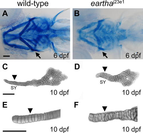
The earthaj23e1 Mutation Affects Cartilage Maturation (A?F) The pharyngeal skeleton of wild-type and earthaj23e1 larvae are revealed by Alcian Blue staining, where the staining in earthaj23e1 larvae (B) is significantly weaker than that in wild-type larvae (A). At 10 dpf, the chondrocytes in the symplectic (SY) region (arrowhead in [C] and [E]) of the hyosymplectic cartilage are arranged into a single cell wide stack (arrowhead in [E]) in wild-type larvae. This organization of chondrocytes is disarranged in the earthaj23e1 larvae (arrowheads in [D] and [F]). Arrows in (A) and (B) indicate the hyosymplectic cartilages that are shown in (C) and (D). Arrowheads in (C) and (D) mark the SY region of hyosymplectic cartilage. (E) and (F) are enlarged images of the SY region of hyosymplectic cartilage. Scale bars: 100 μm.
|