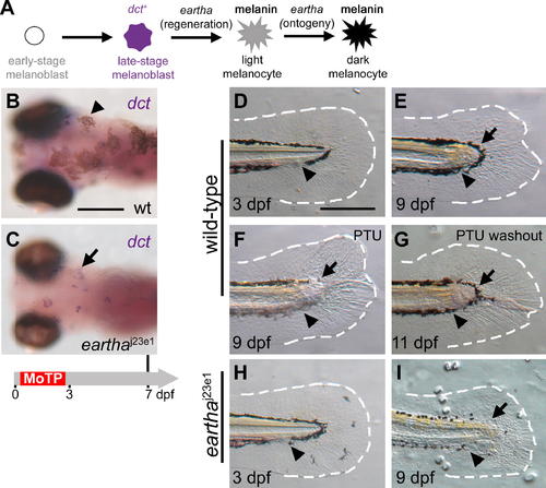
The earthaj23e1 Mutation Specifically Affects Melanocyte Regeneration after the dct+ Stage but Has No Effect on Larval Tail Regeneration (A) The roles of eartha in different stages of melanocyte development in ontogeny and regeneration are presented. (B and C) Larval melanocytes in the wild-type (B) and earthaj23e1 (C) larvae were ablated by MoTP treatment from 14 to 72 hpf, and melanocyte regeneration was analyzed at 7 dpf by ISH with dct riboprobes for late-stage melanoblasts. At this stage, many melanocytes (melanin+) have regenerated in the wild-type larvae (arrowhead in [B]). Although only few pigmented melanocytes appear in earthaj23e1 larvae at the same stage, many dct+ melanoblasts (melanin-, stained purple) are revealed by ISH (arrow in [C]). (D?I) earthaj23e1 mutation specifically affects melanocyte regeneration during larval tail regeneration. Arrowheads show amputation planes at 3 dpf prior to amputation (D) and (H) and after regeneration (E?G) and (I). Wild-type larval tails (D) were amputated at 3 dpf. The wound healed in the ensuing two days by closing the injured tip of the notochord and neural tube, followed by the outgrowth of fin fold. By 9 dpf (6 d postamputation), the missing larval tails were reconstituted, including fin-fold tissues (outlined with white-dashed lines) and melanocytes (arrow in [E]). The origins of the re-established tail melanocytes were investigated by incubating larvae in PTU immediately after larval tail amputations (F). PTU prevents melanin synthesis in newly differentiated melanocytes, and thus the pigmented melanocytes that appear in regenerated tails of the PTU-treated larvae pre-existed prior to the tail amputation. At 9 dpf, few melanocytes (arrow in [F]) appear in the newly regenerated tail immediately distal to the amputation plane in the PTU-treated larvae, suggesting that these melanocytes arise from the migration of proximately differentiated melanocytes from the stump (F). Additional lightly pigmented melanocytes appear after PTU washout (arrow in [G]), indicating that these melanocytes are newly differentiated. When similar tail amputations were conducted in earthaj23e1 larvae (H and I), the larval tails regenerated identically to wild-type larvae, except that few melanocytes appear in the regenerated tail in the region immediately distal to the amputation plane (arrow in [I]). The white-dashed lines outline the fin folds. Timeline (grey) below (C) indicates the period of MoTP treatment (red) and analysis time (vertical line above the timeline). Scale bars: 250 μm.
|

