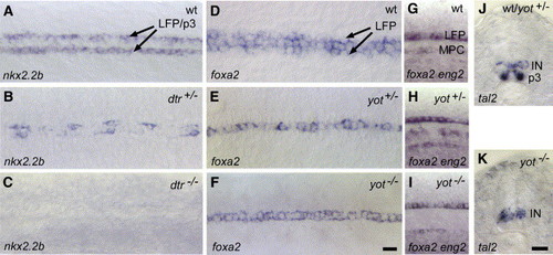
Differential regulation of LFP and p3 neuronal progenitor cells in hedgehog mutants. (A–C) In wild-type control embryos, nkx2.2b is homogenously expressed in LFP and p3 neuronal progenitor cells along the AP axis (A). nkx2.2b expression is reduced to single cells in dtr+/− (detour, gli1) mutants (in n = 15 (56%) out of a clutch of 27 embryos obtained from identified carriers; B) and completely absent in dtr−/− (n = 7/27, 26%; C). Similar results were obtained in yot mutants (yot+/−, n = 22/40, 55%; yot−/−, n = 11/40, 28%). (D) foxa2 is expressed in the MFP and LFP cells in wt embryos (D). In yot+/− (you-too, gli2; E) and yot−/− embryos (F), foxa2 is only detectable in the MFP (n = 23/34, 68%). The same was observed in dtr+/− and dtr−/− mutants (n = 11/18, 61%). (G–I) Identification of homozygous and heterozygous yot mutants. In wild-type embryos, eng2a is expressed in muscle pioneer cells (MPC; G). In yot+/− embryos, where foxa2 expression is lost in the LFP, expression of eng2a is normal (H). yot−/− embryos, in contrast, lack both, foxa2 in the LFP, as well as eng2a in the trunk (I). (J, K) Transverse sections of middle trunk regions. In wt and yot+/− embryos, tal2 is expressed in the p3 neuronal progenitor cells and in dorsally located interneurons (IN) within the ventral neural tube (J). yot−/− embryos lack tal2 expression in p3 neuronal cells, expression in more dorsally located interneurons is unaffected (n = 6/26, 23%; K). Scale bars: F, K, 10 μM.
|

