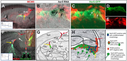
her5-positive cells proliferate and demarcate a new proliferation zone within the adult midbrain. (A-E) Saggital section through a 3-month-old her5:gfp transgenic brain (at the medio-lateral level indicated in A', anterior left), displaying the tectal proliferation zone (red arrow) and the IPZ (yellow arrow) labelled by the cell cycle marker MCM5 (red). her5 RNA-expressing cells (black) and Her5-GFP cells (green) are located in the IPZ. B-E show a higher magnification (single confocal plane) of the section in A showing the expression of the proliferation marker MCM5 (red) in her5-RNA- and Her5-GFP-expressing cells, as pointed by the arrows (B: overlay of bright field and fluorescence views, C: overlay of green and red fluorescence channels; single channels are shown in D and E, respectively). (F) Cross-section through a 3-month-old her5:egfp transgenic brain, at the rostro-caudal level indicated in F′, displaying Her5-GFP (green) and proliferating cells stained by PCNA (red). Yellow arrow to the IPZ, red arrow to the tectal proliferation zone, the location of the connecting ribbon of proliferating cells is indicated by the white arrow. (F″) confocal plane of the area boxed in F showing two Her5-GFP cells that are PCNA-positive (black arrow). (G) Schematic representation of the cross-section shown in F (same colour code for arrows and stainings). (H) Same schematic representation of the IPZ area as in Fig. 1J (sagittal view) depicting the location of PCNA-(or MCM5)-positive cells (red) within the Her5-GFP population. Scale bars: 100 μm in A,F; 10 μm in B. Cb, cerebellum; Ctec, tectal commissure; DTN, dorsal thalamic nucleus; NLV, nucleus lateralis valvulae; PGZ, periventricular grey zone; Teg, tegmentum; TL, torus longitudinalis; TeO, optic tectum; TPZ, tectal proliferation zone; TSc, torus semi-circularis; Val, valvula cerebelli lateralis; Vam, valvula cerebelli medialis.
|

