
Fig. 2
- ID
- ZDB-FIG-060620-2
- Publication
- Villablanca et al., 2006 - Control of cell migration in the zebrafish lateral line: Implication of the gene "Tumour-Associated Calcium Signal Transducer," tacstd
- Other Figures
- All Figure Page
- Back to All Figure Page
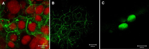
Localization of TACSTD protein. Zebrafish embryos were injected with myc fusion mRNAs, allowed to develop for 12 hr, and developed with an anti-myc antibody. A: Zebrafish tacstd-myc fusion mRNA was injected and embryonic cells were visualized under confocal microscopy; nuclei were labelled with propidium iodide (red) and the anti-myc antibody was detected with an Alexa fluorescent secondary antibody (green). B: Same as in A, but propidium iodide was not added. Note expression localized to membranes and to the perinuclear region. C: Injected mRNA corresponds to myc-tagged SKIP protein, a transcription factor localized to the nucleus. |