
Fig. 4
- ID
- ZDB-FIG-060424-8
- Publication
- Grinblat et al., 1998 - Determination of the zebrafish forebrain: induction and patterning
- Other Figures
- All Figure Page
- Back to All Figure Page
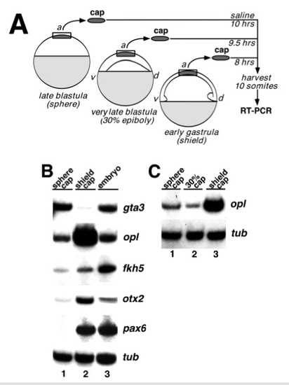
Explant assays show that neural specification occurs by early gastrula. (A) Schematic outline of the explant assay. Animal caps were explanted from sphere stage, 30% epiboly stage,or shield stage embryos. Caps were combined in groups of 5-10 and cultured until control embryos reached mid-somitogenesis, when they were assayed by a reverse transcriptase-PCR (RT-PCR) assay for expression of marker genes, as described in Materials and Methods. (B) RT-PCR analysis of gene expression in animal cap explants. The following markers were assayed: gta3 (Neave et al., 1995), a marker of ventral ectoderm; opl, a marker of prospective telencephalon; fkh5, a marker ofprospective diencephalon, mesencephalon and spinal cord; otx2 (Li et al., 1994), a marker of prospective telencephalon, diencephalon, and mesencephalon; pax6 (Krauss et al., 1991), a marker of prospective diencephalon, hindbrain and spinal cord, and α-tubulin (G. Conway, personal communication), a loading control. Each lane represents a pool of five explants. The data shown here is representative of 2 experiments each for gta3, otx2 and pax6, 3 experiments for fkh5 and 5 experiments for opl. Lane 1: explants from sphere stage embryos; lane 2: explants from shield stage embryos; lane 3: whole embryo controls. (C) RT-PCR analysis of opl expression in animal cap explants from a later blastula stage. Dissections were done as in A using embryos at 30- 35% epiboly. Lane 1: explants from sphere stage embryos; lane 2: explants from 30-35% epiboly stage embryos; lane 3: explants from shield stage embryos. |