Fig. 8
- ID
- ZDB-FIG-060424-12
- Publication
- Grinblat et al., 1998 - Determination of the zebrafish forebrain: induction and patterning
- Other Figures
- All Figure Page
- Back to All Figure Page
|
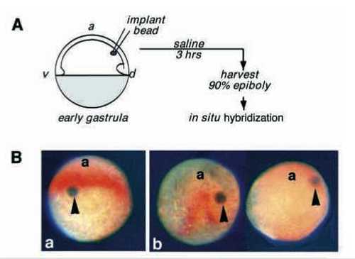
BMP4 inhibits opl expression in whole embryos. (A) Diagram of the experiment. Sepharose beads, coated with BSA or BMP4 (see Methods), were inserted into the dorsal side of shield stage embryos, between the yolk cell and the epiblast. Embryos were harvested at 90% epiboly stage equivalent and assayed for expression of opl (orange) by in situ hybridization. (B) Dorsoanterior views of representative embryos in which control BSA-coated (a) or BMP4- coated (b) Sepharose beads had been implanted. (A) A control embryo with implanted BSA-coated bead (arrowhead). Seven such embryos were obtained in 3 experiments. (b) Embryos obtained after implantation of BMP4 coated beads. 11 embryos generated in 3 experiments had BMP4 beads located dorsally (arrowheads); 3 of these did not express opl (like the embryo on the right), the other 8 showed partial ablation of the opl domain proximal to the bead (like the embryo on the left). a, anterior. |

