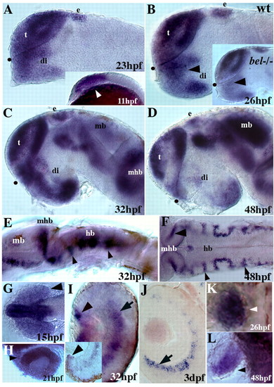
Embryonic expression of zebrafish lhx2. (A) At 23 hpf, lhx2 is expressed in most of the telencephalon, anterior diencephalon and in the epiphysis. Inset shows lhx2 expression in the anterior CNS at the tailbud stage (11 hpf, arrowhead). (B) At 26 hpf, lhx2 is regionally expressed in the telencephalon, diencephalon and epiphysis. Inset shows reduced lhx2 expression only in the dorsal/anterior diencephalon in a bel mutant (arrowhead). (C,D) lhx2 continues to be regionally expressed in the forebrain, midbrain and hindbrain at 32 and 48 hpf. By 48 hpf, lhx2 expression is reduced at the chiasm region and also in the telencephalon. (E) Lateral view shows lhx2 expression in the midbrain/hindbrain border and hindbrain at 32 hpf (arrowheads). (F) Dorsal view shows lhx2 expression at rhombomere boundaries (arrowheads) at 48 hpf. (G) lhx2 expression throughout the eye field (arrowhead) at 15 hpf. Initial expression in the eye field is low relative to that in the forebrain. (H) At 21 hpf, lhx2 is expressed throughout the optic vesicle, with higher levels of expression in the dorsonasal region (arrowhead). (I) At 32 hpf and 48 hpf (inset), lhx2 is expressed in the marginal zones (arrowheads) and in the amacrine cell layer (arrow) of the eye. (J) By 3 dpf, expression in the amacrine layer becomes restricted ventrally (arrow). (K,L) lhx2 is expressed in most of the fin bud at 26 hpf (K) and becomes restricted to the posterior bud at 48 hpf (L, arrowheads). (A-E) Lateral views, anterior to the left; (F,G,J,K) dorsal views; (H,I) 7 μm sections of the eye. Black dots mark the position of the optic recess. e, epiphysis; di, diencephalon; hb, hindbrain; mb, midbrain; mhb, midbrain-hindbrain boundary; t, telencephalon.
|

