Fig. 1
- ID
- ZDB-FIG-060208-1
- Publication
- Seth et al., 2006 - belladonna/(lhx2) is required for neural patterning and midline axon guidance in the zebrafish forebrain
- Other Figures
- All Figure Page
- Back to All Figure Page
|
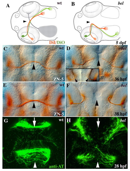
Retinal and commissural axon defects in bel. (A) Schematic showing that all retinal axons cross the wild-type midline (arrowhead) and project topographically on the contralateral tectal lobe [anterior/dorsal retina (green) to posterior/ventral tectum, posterior/ventral retina (red) to anterior/dorsal tectum]. (B) In bel mutants, retinal axons fail to cross the midline (arrowhead) and instead project to the ipsilateral tectal lobe in a topographically correct manner (see Karlstrom et al., 1996). (C) At 36 hpf, wild-type RGC axons have reached the midline (arrowhead). (D) In most bel mutants, RGC axons have grown towards the midline (arrowhead), but in some mutants axons project ipsilaterally immediately after leaving the eye (inset, arrowheads). (E) At 38 hpf, wild-type RGC axons have crossed the midline (arrowhead). (F) In bel mutants, all RGCs project ipsilaterally at 38 hpf. (G) Both the AC (arrow) and POC (arrowhead) are fully formed in wild-type embryos by 28 hpf. (H) In bel mutants, both forebrain commissures fail to form. (A,B) Schematic dorsal views of the head, anterior left (adapted, with permission, from Culverwell and Karlstrom, 2002). (C-H) Ventral views of the forebrain, eyes on either side of each panel, anterior up. |

