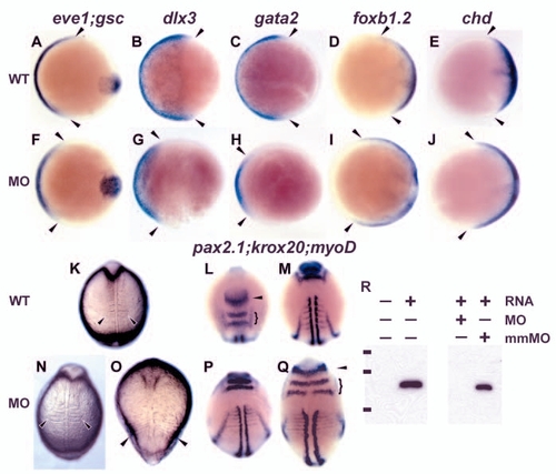
High level tsg1 knockdown dorsalizes the embryo. Uninjected embryos (A-E,K-M) compared with siblings injected with 25 ng MO5 (F-J,N,P,Q) or 32 ng MO1 (O). (A-J) Whole-mount in situ hybridization at 80% epiboly (mid-gastrulation); animal pole views, dorsal towards the right. Reduced ventral domain of eve1 (n=32/54), dlx3 (n=25/41) and gata2 (n=28/50) expression (A-C,F-H) and expanded foxb1.2 (n=34/46) and chordin (chd) (n=16/30) expression in dorsal regions (D,E,I,J) in injected compared with uninjected embryos (delineated by arrowheads) was observed. gsc expression in the dorsal midline prechordal plate mesoderm is not affected (A,F), similar to results in other dorsalized BMP signaling pathway mutants in zebrafish (Mullins et al., 1996; Nguyen et al., 1998). (K-Q) During somitogenesis, injected embryos display dorsalized characteristics similar to class 3 (N) and class 4 (O) dorsalized mutants, as assessed by the lateral extent of somites in live embryos (arrowheads in K,N,O), by whole-mount in situ hybridization with myod in anterior somitic mesoderm (M,P,Q), and by expansion of pax2.1 (arrowhead) and krox20 (bracket) expression in the MHB and rhombomeres 3 and 5, respectively (L,Q). The embryos in N,P display a class 3 or moderate dorsalization, whereas those in O,Q exhibit a greater expansion of the somitic mesoderm and neural tissue, similar to a class 4 dorsalization. (L) Dorsoanterior view; (M) a more posterior view of the same uninjected embryo. (R) Anti-FLAG western blot on lysates of embryos injected with FLAG-tagged tsg1 alone or with 32 ng MO1 or mismatch MO1. Bars to the left of the blot represent the positions of protein standards with molecular weights of 50, 37 and 25 kDa.
|

