|
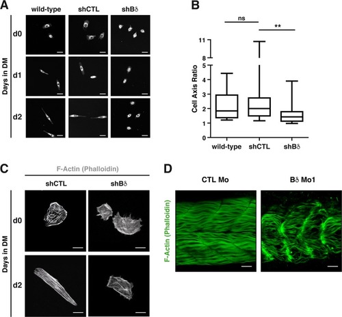
PP2A-Bδ is involved in fusion-associated cytoskeleton morphogenesis.a Representative (n = 2) confocal images of morphology of wild-type (WT), control (shCTL), and Bδ-knocked down (shBδ) C2C12 myoblasts at the indicated time points during the differentiation process. Cells were stained with CellMask (white). Scale bars are 25 µm. b Major/minor cell axis ratio in wild-type (n = 21), control (shCTL, n = 29), and Bδ-knocked down (shBδ, n = 27) C2C12 myoblasts at day 2 during the differentiation process. Kruskal–Wallis with Dunn’s correction, *P < 0.05, **P < 0.01. ns: not significant. c Confocal analysis of F-actin (phalloidin, gray) in control (shCTL) and Bδ-knocked down (shBδ) C2C12 myoblasts grown in GM (d0) or 2 days in DM (d2). Representative images from two experiments are shown. Scale bars are 10 µm. d Representative (n = 3 experiments) confocal imaging of F-actin (phalloidin, green) in 48-hpf control (CTL Mo) or PP2A-Bδ (Bδ Mo1) morphant embryos. Scale bars are 25 µm
|