|
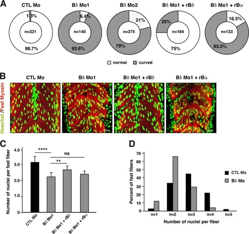
PP2A-Bδ holoenzyme is necessary for myofiber formation <italic>in vivo</italic>.a, b Quantification of the curved trunk phenotype (a) and b representative confocal imaging in 48-hpf zebrafish embryos injected with a control morpholino (CTL Mo), with an ATG-blocking morpholino alone (Bδ Mo1) or together with a rat Bδ (Bδ Mo1 + rBδ) or rat Bα mRNA (Bδ Mo1 + rBα) or an alternative splice-blocking morpholino (Bδ Mo2) against PP2A-Bδ. Nuclei (Hoechst) and fast skeletal myosin are shown respectively in green and red. Scale bars are 25 µm. c Quantification of the number of nuclei in fast skeletal myofibers in 48-hpf control embryos (CTL Mo, n = 16) or in embryos injected with the PP2A-Bδ Mo alone (Bδ Mo, n = 20) or together with a rat Bδ (Bδ Mo + rBδ, n = 7) or rat Bα mRNA (Bδ Mo1 + rBα, n = 9). Values are mean ± SD, unpaired t-test, two-tailed, ****P < 0.0001. d Proportion of fibers with the indicated number of nuclei in control (CTL Mo, n = 11) or PP2A-Bδ (Bδ Mo, n = 14) morphant embryos
|