
- Title
-
The Risk Genes for Neuropsychiatric Disorders negr1 and opcml Are Expressed throughout Zebrafish Brain Development
- Authors
- Habicher, J., Sanvido, I., B�hler, A., Sartori, S., Piccoli, G., Carl, M.
- Source
- Full text @ Genes (Basel)
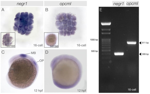
EXPRESSION / LABELING:
|
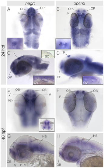
Expression patterns of |
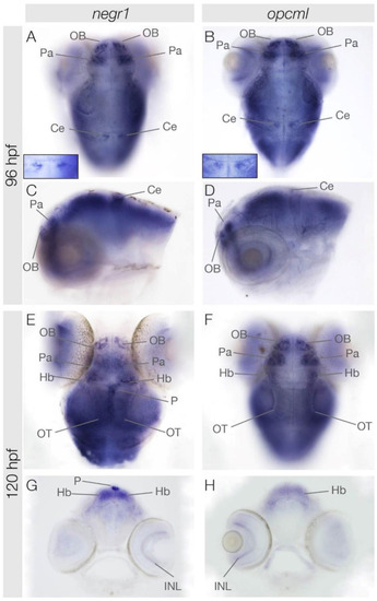
Expression patterns of |