FIGURE SUMMARY
Title

Iroquois transcription factor irx2a is required for multiciliated and transporter cell fate decisions during zebrafish pronephros development

Authors
Marra, A.N., Cheng, C.N., Adeeb, B., Addiego, A., Wesselman, H.M., Chambers, B.E., Chambers, J.M., Wingert, R.A.
Source
Full text @ Sci. Rep.

irx2a expression localizes to a region of the zebrafish pronephros that corresponds to the PST, DE and MCC domains. (A) Schematic of a zebrafish embryo (lateral view) at 24 hpf, which is equivalent to the 28 ss. Schematic below depicts color coded segments, corresponding somite numbers, and the expression pattern of MCCs in black within the nephron. MCC number is not to scale. (B) WISH in wild-type zebrafish embryos at the 15–18 ss, 20–22 ss, and 28 ss demonstrates irx2a transcript expression (purple) in the middle of the developing pronephros. Black lines highlight the irx2a expression domain. Scale bar is 50 μm. (C) FISH in wild-type embryos at the 28 ss demonstrates that irx2a expression (magenta) colocalizes with cdh17+ epithelial cells and (D) odf3b+ MCCs of the nephron tubule (green), where DAPI labels the nuclei (grey). Scale bar is 50 μm. White circles indicate nuclei with co-expression of the respective markers. P - podocytes, N - neck, PCT - proximal convoluted tubule, PST - proximal straight tubule, DE - distal early, DL - distal late, MCC - multiciliated cell, ss - somite stage, WISH – whole mount in situ hybridization, FISH – fluorescent in situ hybridization.

irx2a restricts the PST and promotes the DL during pronephros segmentation. (A) Lateral view of WISH analysis with the PST marker trpm7 at the 28 ss. Black bars highlight the trpm7 expression domain in the pronephros. Scale bar is 50 μm. (B) Quantification of trpm7 length (μm). Each dot represents one nephron and data is presented +/− SEM. Statistical significance was determined with ANOVA. (C) WISH in 28 ss embryos with the DL marker slc12a3 shown in a lateral view. Black bars denote slc12a3 expression. (D) Quantification of slc12a3 domain length (μm), where each dot represents one nephron. Data is presented +/− SEM and statistical significance was determined by ANOVA. WISH – whole mount in situ hybridization, ss – somite stage, PST – proximal straight tubule, DL – distal late tubule.

irx2a acts upstream of etv5a. WISH at the 28 ss revealed decreased etv5a expression in the pronephros of irx2a morphants. Black bars denote the etv5a expression domain. Scale bar is 50 μm. (B) Quantification of etv5a length in μm at the 28 ss. Each dot represents one nephron and data is presented +/− SEM. Significance was evaluated with an unpaired student’s T-test. (C) The irx2a domain is unchanged in etv5a morphants at the 28 ss. Solid black bars denote strong irx2a expression and the dashed black lines denote faint expression in the pronephros. (D) Quantification of irx2a length (μm) at the 28 ss. Each dot represents one nephron and data is presented +/− SEM. An unpaired student’s T-test was used to determine significance. WISH – whole mount in situ hybridization, ss – somite stage.

EXPRESSION / LABELING:
Genes:
Fish:
Knockdown Reagents:
Anatomical Term:
Stage: Prim-5
PHENOTYPE:
Fish:
Knockdown Reagent:
Observed In:
Stage: Prim-5

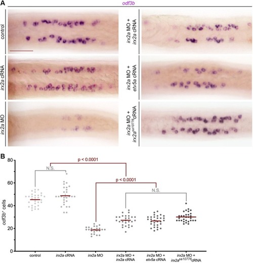
irx2a is required upstream of etv5a for MCC genesis. (A) Dorsal view of odf3b in the pronephros by WISH at the 28 ss shows a decrease in maturing MCCs in irx2a deficient embryos, which was rescued with irx2a cRNA, etv5a cRNA, and irx2asa10776 cRNA. Scale bar is 50 μm. (B) Quantification of odf3b+ MCCs at the 28 ss. Each dot represents one pronephros. Data is presented +/− SEM and statistical significance was determined with ANOVA. WISH – whole mount in situ hybridization, ss – somite stage, MCC – multiciliated cell.

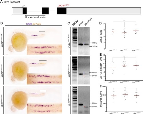
irx2a genetic mutants do not have a renal or eye phenotype. (A) Schematic of the irx2a transcript and location of the irx2asa10716 mutation, where grey is non-coding and black represents coding sequence. The forward arrow demarcates the ATG start site. (B) WISH analysis at the 28 ss shows odf3b and slc12a3 expression in irx2asa10716+/+, irx2asa10716+/−, and irx2asa10716−/− embryos. The black lines highlight the DL marker slc12a3. An enlarged dorsal view of the odf3b+ MCCs for each embryo is shown in the inset. Scale bars are 50 μm. (C) Gel image of the restriction digest product for each genotype. Wild-type bands are marked with a blue star, and mutant bands with a red star. (D) Quantification of odf3b+ cells at the 28 ss. Each dot represents one pronephros. (E) Quantification of the slc12a3 length (μm) at the 28 ss. Each dot represents one nephron. (F) Quantification of eye area (μm2). Each dot represents one eye. For all graphs, data is presented +/− SEM and statistical significance was determined by ANOVA. WISH – whole mount in situ hybridization, ss – somite stage, DL – distal late, MCC – multiciliated cell.

RA signaling regulates irx2a expression during nephrogenesis. (A) WISH analysis at the 28 ss demonstrates alterations in the irx2a pronephros domain after either treatment of RA or DEAB. The black arrowheads show the distance from distal irx2a expression to the cloaca, and the black lines highlight irx2a expression. (B) Quantification of irx2a length (μm) at the 28 ss. Each dot represents one pronephros. (C) Quantification of the length from irx2a expression to the cloaca (μm), where each dot represents one pronephros. For each graph, data is represented +/− SEM and significance was determined with an ANOVA. WISH – whole mount in situ hybridization, ss- somite stage, RA – retinoic acid, DEAB - N,N-diethylaminobenzaldehyde.

Acknowledgments
This image is the copyrighted work of the attributed author or publisher, and ZFIN has permission only to display this image to its users. Additional permissions should be obtained from the applicable author or publisher of the image. Full text @ Sci. Rep.