- Title
-
Kinesin-1 promotes chondrocyte maintenance during skeletal morphogenesis
- Authors
- Santos-Ledo, A., Garcia-Macia, M., Campbell, P.D., Gronska, M., Marlow, F.L.
- Source
- Full text @ PLoS Genet.
|
kif5Bs contribute to craniofacial development. A) Using Crispr-CAS9 mutagenesis to target the N-terminus, ae21 (9bp deletion), ae22 (4bp deletion), ae23 (5bp deletion) and ae24 (2bp insertion causing a frame shift and early stop codon) kif5Bb mutant alleles were generated. Exon is indicated in yellow, PAM region in orange, red dash or blue areas indicate mutated regions. B) qRT-PCR for kif5Ba and kif5Bb in kif5Baae12/ae12 and kif5Bbae24/ae24 embryos. kif5Bb transcripts undergo NMD in kif5Bbae24/ae24 mutants. One-way ANOVA * p<0.05. C-E) Live images of 6dpf kif5Bs mutants. Mutation of kif5Ba causes incompletely penetrant jaw protrusion defects (arrow in D and Dī). kif5Baae12/ae12Kif5Bbae24/ae24 mutants exhibit a fully penetrant phenotype, including flat heads (arrows in E and Eī). F) Measurements of the jaw protrusion in each genotype. One-way ANOVA ** p<0.01, *** p<0.001, **** p<0.0001. G-J) Whole-mount in situ hybridization of sox9a (G, H) and col2a1 (I, J) revealed no differences between Wt and mutants. Arrows indicate the cartilage region. K-N) Alcian blue staining at 3 dpf, arrow indicates inset region. Intact cartilage elements in kif5blof are shorter and mediolaterally broader, the angle at the ceratohyal cartilage intersection is wider (arrowheads in K, N). Alcian blue stain reveals defective chondrocyte stacking (insets in K-N). O-R) Alcian blue staining at 5 dpf, arrow indicates the inset region. Meckelīs cartilage has extended anteriorly (O) in Wt but not in kif5Blof mutants. (P-R) the Meckelīs and ceratohyal cartilages are curved in kif5Blof (arrowheads in O-R), Alcian blue staining is diffuse, and cell stacking is absent (insets in P-R). ch: ceratohyal; m: Meckelīs; pq: palatoquadrate. Scale bars: A-H: 500?m; I-P: 50?m. EXPRESSION / LABELING:
PHENOTYPE:
|
|
Loss of kif5B impairs chondrocyte secretion. A-C) confocal images of Wt (A), single kif5Baae12/ae12 mutants (B) and kif5Baae12/ae12Kif5Bbae24/ae24 (C) at 60 hpf. Chondrocytes (Sox10:GFP positive cells) secrete proteins positive for peanut agglutinin (PNA, blue) homogeneously. Secretion in single (B) and compound (C) mutants is deficient. Stacking defects are evident in kif5Baae12/ae12Kif5Bbae24/ae24 (C). D-F) Confocal images of Wt (D), single kif5Baae12/ae12 mutants (E) and kif5Baae12/ae12Kif5Bbae24/ae24 (F) at 5 dpf. In Wt (D) Di0C6 (blue) forms variably-sized cellular aggregates. Wheat Germ agglutinin (WGA, green) labels secreted proteins and II-II6B3 marks Collagen II (red) an abundant protein in cartilage. Both are homogeneously secreted in Wt and overlap within ECM (insets in D), although some overlap occurs between Collagen II and Di0C6 (insets in D). In single (E) and compound (F) mutants, Di0C6 forms large patches. Secretion is polarized with little colocalization within ECM (insets in E and F), large patches of Di0C6 colocalize with Collagen II, and cell stacking is perturbed (E, F). Scale bar: 50?m. |
|
mpeded mineralization in kif5Blof. Confocal projections of images of live embryos at 6 dpf (A, D) and 11 dpf (B, C, E, F). GFP stained chondrocytes and Alizarin Red stained mineralized matrix. Intramembranous bones (MD, P and B) show mineralization but were shorter at 6 dpf (A, D). BC, the only endochondral bone at this stage, mineralization was deficient but evidence of mineralization is detectable (insets in D). At 11 dpf membranous and perichondral ossification had progressed in Wt (B) and vertebraes were apparent (C). In kif5Blof membranous ossification had also progressed (E) and vertebraes were mineralized (F). However, perichondral ossification was defective as indicated by the absence of BC (E). B: branchiostegal; BC: bone collars; P: entopterygoid; D: dentary. |
|
kif5Blof defects and interactions with Wnt PCP components. A-F) Semi-thin sections of the trabecular (A, B) and palatoquadrate cartilage (C-F). No differences were detected at 48 hpf (A, B) or 60 hpf (C, D). At 3 dpf, in Wt (E) chondrocytes stack like coins and span the cartilage. However, kif5Blof chondrocytes are circular (F), and close to the cartilage border. Some cells are delineated with dashed outlines and in E-F the cartilage limit is drawn. G-I) Analysis of MTOC orientation using ?-Tubulin. In Wt embryos (G, Gī) >80% were anteriorly oriented (I). MTOC position was random in kif5Blof mutants (H, H'). WT: n = 8; 72cells. kif5Blof: n = 5; 63 cells (I). J-K''') Confocal images of single kny (J-J''') or triple mutants (K-K''') at 5 dpf. Secretion is normal in single kny mutants (J'-J''') but stacking is defective (J'''). Stacking and secretion defects in triple mutants (K-K'''). L-M''') Confocal images of single ppt (L-L''') or triple mutants (M-M'''). Secretion is intact in ppt single mutants (L'-L''') but stacking is disrupted (L'''). Membrane (M), secretion (M', M'') and stacking (M''') deficits are evident in triple mutants. ch: ceratohyal; m: Meckelīs; pq: palatoquadrate. Scale bars: A-F: 50?m; G, H: 20?m, J-M''': 50?m. EXPRESSION / LABELING:
PHENOTYPE:
|
|
Autophagy and chondrocyte maintenance in kif5Blof cartilage. A-D) Electron microscopy of the Wt palatoquadrate. At 3 dpf Wt rER and mitochondria are apparent within stacked chondrocytes (A, B and inset). In the proliferative region (PR), cells are round (C). Cartilage is dense near the perichondrium (arrow in A). At 5 dpf, Wt chondrocytes are hypertrophic with large vacuoles but are stacked (D). E-H) At 3 dpf, kif5Blof cartilage contains small, round highly vacuolated cells (E, F and inset) with pronounced rER expansion (E and inset). Both rER and vacuoles appear to contain cellular components (F and inset in E). In the PR, overall chondrocyte morphology is more normal, but with the previously mentioned intracellular characteristics (G). Nuclei are patchy and distributed towards the cartilage border (E, F). Cells are extruded from the cartilage (arrow in E) and the region near the perichondrium is disorganized (arrowhead in E). At 5 dpf, degeneration persists and cartilage ?ghosts? remain (arrow in H). I, J, K) Tunel (apoptotic) cells were rare in wild-type embryos, but are abundant in kif5Blof mutants. Tunel-positive cells in the ceratohyal (10 embryos were quantified/group). L) ER stress marker (bip1 and sil1) levels. Scale bar: A, D, E, H: 2?m; B, C, F, G: 1?m. n.s. no significant; *** p<0.001 (Student T test). M-S) At 4 dpf in basal conditions, Wt embryos show LC3 positive puncta in chondrocytes (M), which increases after exposure to NH4Cl (N). Basal LC3 staining is higher in kif5Blof mutants (O) and increases less after drug exposure (P). LC3 fluorescence intensity quantification (5?10 cells in at least 10 embryos/group) (Q, ANOVA). LC3 flux as measured by western blot in embryos with normal jaws and with short jaws (R, LC3 protein and Ponceau staining to reveal total protein loaded, S, quantification, Student T test). I, J) Lysotracker (magenta) stains acidic cellular compartments, including lysosomes. In Wt chondrocytes, several puncta are observed near the nucleus (T). Lysotracker accumulation in kif5Blof mutants (U). V) Reduced Tor activation in embryos with short jaws (V, P-Tor and Tor western blot and Ponceau staining to assess total protein loaded, Student T test, n = 4). W-Y) Phospho-S6 Ribosomal Protein (PS6) abundance in chondrocytes of Wt embryos (W) and in kif5blof mutants (X), quantified in Y (At least 5?10 cells in at least 10 embryos, Student T test). NJ: normal jaw (Wt), SJ: short jaw (kif5Baae12/ae12Kif5Bbae24/+ and kif5Baae12/ae12Kif5Bbae24/ae24). EXPRESSION / LABELING:
PHENOTYPE:
|
|
Cell autonomous and cell-nonautonomous kif5B effects. A) Cartoon depicts the experimental design. B-F) Confocal images of sectioned palatoquadrate cartilage at 3 dpf. WT cells (GFP+) transplanted to Wt embryos integrate and secrete matrix (B). Single Wt cells (GFP+) in kif5Blof elongate, secrete and stack (arrows in C), and the nucleus is centrally positioned compared to mutants (arrowhead in C). Several Wt cells (GFP+) rescue cell stacking in nearby mutant neighbors (arrows in D). Rescue correlates with strong WGA expression along the cartilage border (arrowheads in D). Defects of single kif5Blof cells (GFP+) persist in Wt hosts (E) including secretion (arrows in E) and nuclear position (arrowhead in E), although the cells partially elongate. Groups of kif5Blof cells (GFP+) in Wt hosts (F) did not stack or elongate and had secretion deficits. Cells are extruded from the cartilage (arrows in F). Successful transplants: Wt to Wt: 13; Wt to kif5Baae12/ae12kif5Bb+/+: 2; Wt to kif5Baae12/ae12kif5Bbae24/+: 8; Wt to kif5Baae12/ae12kif5Bbae24/ae24: 2; kif5Baae12/ae12kif5Bb+/+ to Wt: 6; kif5Baae12/ae12kif5Bbae24/+ to Wt: 3; kif5Baae12/ae12kif5Bbae24/ae24 to Wt: 0. In the figure, the numbers indicate transplants with similar scenarios for each genotype. Scale bar: 20?m. |
|
Rescue of kif5Blof by Kif5Ba but not Kif5Aa in cartilage. A) Cartoon depicts experimental design. B-E) Kif5Ba or Kif5AaOE did not cause phenotypes in Wt (B, D). Kif5BaOE in kif5Blof mutants rescues the phenotype (C, compare right to left). Chondrocytes on the rescued side were stacked and elongated with proper secretion. Rescue improved when both chondrocytes and perichondrium expressed kif5Ba (arrows in C) versus chondrocytes alone (arrowheads in C). Overexpression of the neural specific kif5Aa did not rescue kif5Blof phenotypes (E). |
|
Muscle phenotypes of kif5Blof mutants. A-D) Ventral view of the anterior region of zebrafish at 5 dpf. Muscle fibers are shorter than Wt, and broken muscle fibers are detected along the body axis (arrows in C and D). This phenotype was not observed in kif5Ba single mutants (B). E-H) Lateral views of the tail at 5 dpf; broken muscle fibers are observed in kif5Blof mutants (arrows in G and H). I-L) Electron microscopy of the ocular muscle at 3 dpf (I, J) and 5 dpf (K, L). No disruption of muscle ultrastructure was apparent at 3 dpf (I, J). However, at 5 dpf the M-line of the sarcomere is diminished in kif5Blof mutants (I, J and insets). PHENOTYPE:
|
|
Central nervous system is not altered in kif5Blof mutants. Ventral (A, D) or lateral (B, E) view of the head and lateral view of the tail (C, F) of Acetylated tubulin staining in Wt (A-C) and kif5Blof mutants (D-F). |
|
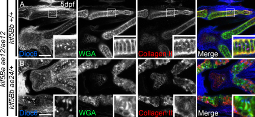
Secretion defects in kif5B compound mutants. Some kif5B single mutants (kif5Baae12/ae12Kif5Bb+/+) resembled Wt in terms of membrane distribution and secretion (A). kif5B compound mutant (kif5Baae12/ae12Kif5Bbae24/+) phenotypes were fully penetrant and indistinguishable from double mutants. |
|
MMP14 distribution is not altered in kif5Blof mutants. Chondrocytes and perichondrium are positive for MMP14 in Wt (A) and kif5Blof (B). Western blot reveals no significant changes [90] in MMP14 levels (C). Scale bar: 1 ?m. D-G) Wt embryos show no kif5Blof-like phenotypes when exposed to broad-spectrum metalloproteinase inhibitor (E, F) or the MMP inhibitor GM6001 (G), although some cells were extruded from the cartilage at high concentrations of EDTA (arrow in F). |


