- Title
-
The chaperone activity of 4PBA ameliorates the skeletal phenotype of Chihuahua, a zebrafish model for dominant osteogenesis imperfecta
- Authors
- Gioia, R., Tonelli, F., Ceppi, I., Biggiogera, M., Leikin, S., Fisher, S., Tenedini, E., Yorgan, T.A., Schinke, T., Tian, K., Schwartz, J.M., Forte, F., Wagener, R., Villani, S., Rossi, A., Forlino, A.
- Source
- Full text @ Hum. Mol. Genet.
|
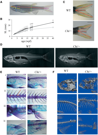
Morphological characterization of adult (Chi/+) zebrafish. (A) Morphometric parameters measured in adult fish. Standard length (SL) in red, height at anterior of anal fin (HAA) in green, height at eye (HE) in blue, snout-operculum length (SOL) in yellow, major axes of the caudal fin in purple, minor axis of the caudal fin in pink. (B) The growth curve shows that mutant fish (n?=?15, grey triangles) are significantly smaller than WT (n?=?12, black squares) from the 18th week post-fertilization (wpf). SL: standard length. *P?<?0.05. (C) Representative image of WT and Chi/+?caudal fin. The caudal fin of mutant fish has less pronounced lobes than the WT fin. The distances measured as described in Materials and Methods are highlighted in red. Scale bar: 1?mm. (D) WT and Chi/+?X-rays show severe alterations in the skeleton of mutant fish. (E) Skeleton magnifications of 3?months old WT and Chi/+?fish stained with alcian blue and alizarin red. Major and minor axes of the vertebral body and the slope of the neural spine are indicated (ii). Deformed ribs with bone calli (i), fused vertebrae (ii, arrow), misshapen prezygapophysis and post-zygapophysis (ii, white and black arrowheads), deformed pectoral (iii) and pelvic fins (iv) are evident in mutant fish. Scale bar: 500 Ám. (F) ÁCT of 3?months old WT and Chi/+?fish reveals the severe under mineralization of mutant animals. PHENOTYPE:
|
|
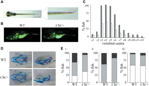
Skeletal evaluation of Chi/+?larvae. (A) Fin fold angle is represented in red in 11 dpf fish. Scale bar: 1?mm. (B) Representative images of 5 dpf larvae WT and Chi/+?stained with calcein. The delay in vertebral centra ossification is evident. Scale bar: 250 Ám. (C) Percentage of 11 dpf fish presenting calcein positive (mineralized) vertebrae. c: vertebral centrum. Grey: WT; white: Chi/+. (D) Alcian blue and alizarin red staining of WT and Chi/+?11 dpf larvae in ventral and lateral orientation. 5th ceratobranchyal (5CB, arrowhead), cleithrum (CL, arrow) and notochord (NC, asterisk) are indicated. Scale bar: 200 Ám. Delayed mineralization, represented by reduced alizarin red staining, is evident in the mutant larvae. (E) Percentage of 11 dpf WT and Chi/+?larvae presenting distinctive levels of mineralization in the 5CB (i), the CL (ii) and the NC (iii). White indicates beginning/no mineralization, grey indicates incomplete mineralization and black indicates complete mineralization. |
|
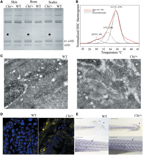
Biochemical and cellular evaluation of Chi/+?collagen type I. (A) SDS-PAGE of collagen type I extracted from skin, bone and scales of WT and Chi/+?adult fish. Mutant collagen is characterized by broader ? bands (asterisks), indicating collagen type I overmodification. (B) Representative DSC thermogram of collagen extracted from bone of Chi/+?adult fish. The presence of three melting temperature (Tm) peaks is evident from the deconvolution analysis of the thermogram. Similar result was obtained for collagen extracted from skin and scales (data not shown). (C) Transmission electron microscopy image of the caudal fin of WT and Chi/+?adult fish. The enlargement of endoplasmic reticulum (ER) cisternae is evident in mutant fish. N: nucleus; *: rough endoplasmic reticulum (ER). Magnification 12000X. (D) Confocal microscopy images of 24 hpf embryos WT and Chi/+?injected with mRNA expressing yellow fluorescent protein in the ER. A stronger signal in the mutant sample is demonstrated. Magnification 40X, zoom 4X. (E) Whole mount immunohistochemistry of 5 dpf WT and Chi/+?embryos using Hsp47b antibody. A stronger signal is evident in tail, skin and intersomitic space in mutant larvae. Arrow: intersomitic space, arrowhead: tail; asterisk: skin. Scale bar: 200 Ám. PHENOTYPE:
|

ZFIN is incorporating published figure images and captions as part of an ongoing project. Figures from some publications have not yet been curated, or are not available for display because of copyright restrictions. PHENOTYPE:
|
|
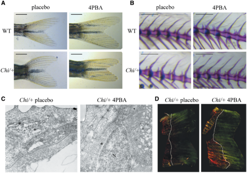
4PBA treatment ameliorated the tail shape (A) and normalized the neural spine slope (B) of Chi/+?fish after 14?weeks of treatment. Scale bar: 1?mm in (A) and 500 Ám in (B). (C) Transmission electron microscopy of 11 dpf Chi/+?larvae showed a general reduction of the ER cisternae size after 4PBA treatment. Magnification 12000X. (D) Representative images of picro sirius red staining Chi/+?zebrafish regrown tail after 7 days from tail clipping in absence or presence of 4PBA. A strongest signal in the sample obtained from 4PBA treated fish confirmed a higher amount of collagen in the matrix following 4PBA treatment. White lines indicate the portion of tail regrown in 7 days. Magnification 10X. PHENOTYPE:
|


