
|
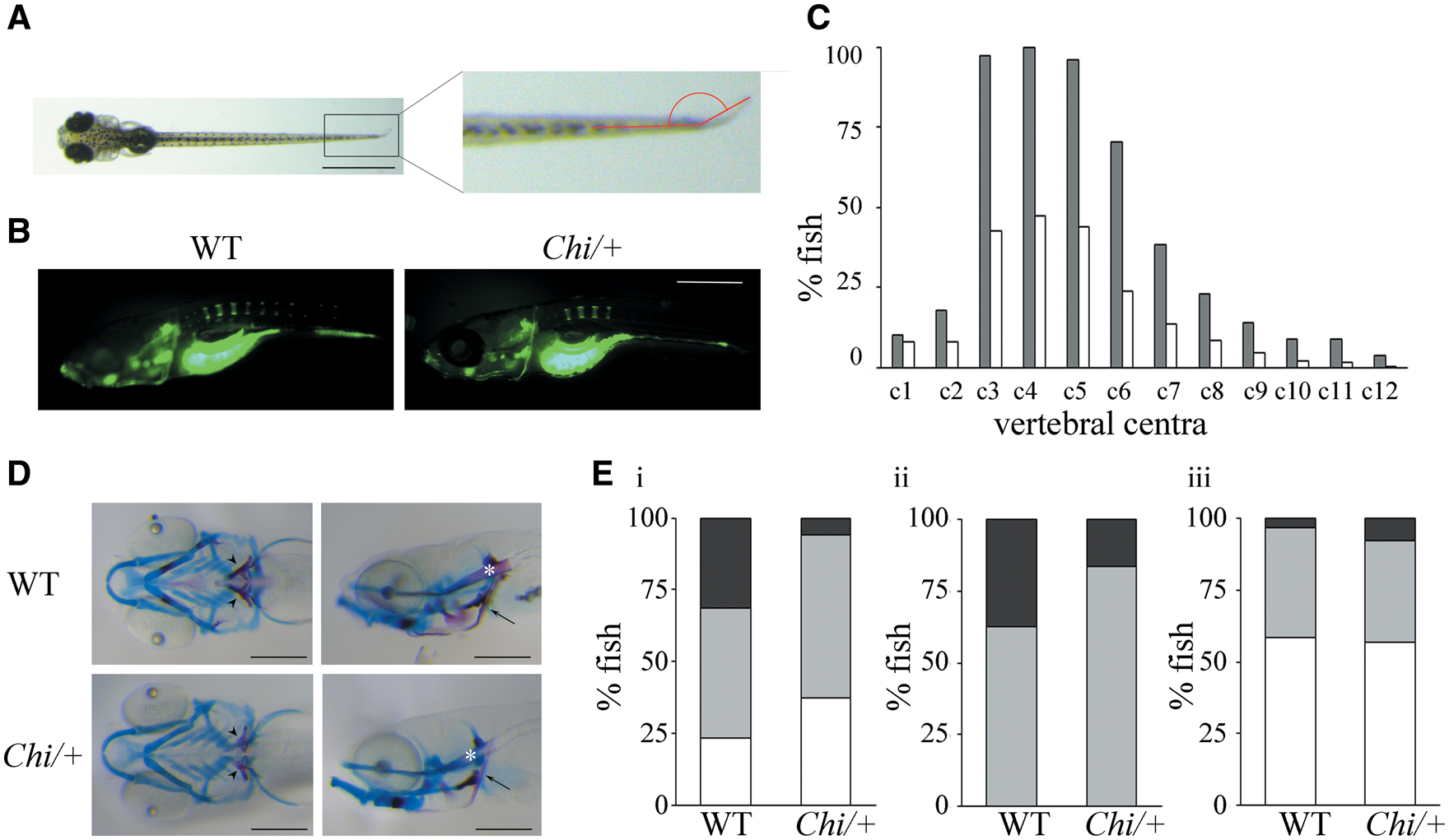
Fig. 2
Skeletal evaluation of Chi/+ larvae. (A) Fin fold angle is represented in red in 11 dpf fish. Scale bar: 1 mm. (B) Representative images of 5 dpf larvae WT and Chi/+ stained with calcein. The delay in vertebral centra ossification is evident. Scale bar: 250 µm. (C) Percentage of 11 dpf fish presenting calcein positive (mineralized) vertebrae. c: vertebral centrum. Grey: WT; white: Chi/+. (D) Alcian blue and alizarin red staining of WT and Chi/+ 11 dpf larvae in ventral and lateral orientation. 5th ceratobranchyal (5CB, arrowhead), cleithrum (CL, arrow) and notochord (NC, asterisk) are indicated. Scale bar: 200 µm. Delayed mineralization, represented by reduced alizarin red staining, is evident in the mutant larvae. (E) Percentage of 11 dpf WT and Chi/+ larvae presenting distinctive levels of mineralization in the 5CB (i), the CL (ii) and the NC (iii). White indicates beginning/no mineralization, grey indicates incomplete mineralization and black indicates complete mineralization.