- Title
-
Disruption of pdgfra alters endocardial and myocardial fusion during zebrafish cardiac assembly.
- Authors
- El-Rass, S., Eisa-Beygi, S., Khong, E., Brand-Arzamendi, K., Mauro, A., Zhang, H., Clark, K.J., Ekker, S.C., Wen, X.Y.
- Source
- Full text @ Biol. Open
|
RP2 Transposon integration disrupts pdgfra gene. (A) Bright field (left) and fluorescent (right) images of heterozygous GBT1300 larva showing mRFP expression pattern at 96?hpf. (B) Schematic representation of zebrafish pdgfra gene showing the locus of RP2 insertion in intron 16, and the resulting truncated pdgfra-mRFP and GFP-pdgfra fusion transcripts. Gray boxes indicate exons; lines indicate introns. Primers used for reverse transcription (RT)- and quantitative real-time (qRT)-PCR are denoted. (C) Schematic drawing of zebrafish Pdgfra protein domains, showing the site of mRFP fusion (red arrow). IG, immunoglobulin; TM, transmembrane; TK, tyrosine kinase. Scale bar: 500??m in A. EXPRESSION / LABELING:
|
|
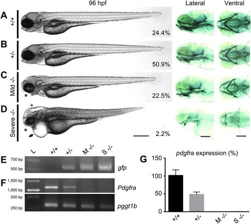
General phenotype analyses in pdgfra loss-of-function mutants. Panels showing bright field (left), and Alcian Blue-stained cartilaginous tissue (right) of WT (+/+) (A), heterozygous (+/?) (B), mild homozygous (Mild/M ?/?) (C), and severe homozygous (Severe/S ?/?) (D) GBT1300 siblings at 96?hpf. The percentage of fish in each group is indicated (n=275). While +/+ and +/? larvae show no visible abnormalities, mild ?/? mutants exhibit craniofacial defects (asterisk) and abnormal swim bladder inflation. Severe ?/? mutants demonstrate severe developmental defects, including craniofacial defects (asterisk), pericardial edema (arrow), hydrocephalus (cross), smaller body axis elongation, and abnormal cardiac and skeletal muscle. (E) PCR indicating the presence of gfp in +/?, mild ?/? and severe ?/?, but not in +/+ fish. (F) RT-PCR demonstrating reduction in endogenous pdgfra transcripts in mild ?/? and severe ?/?, compared to +/+ and +/? fish at 96?hpf. pggt1b used as a control for RT-PCR. (G) Relative amounts of pdgfra transcript confirming the loss of WT pdgfra transcripts in both mild ?/? and severe ?/? mutants at 96?hpf through qRT-RCR (P?0.01; n=25/group; unpaired t test, error bars indicate s.e.m.). Scale bar: 250??m. Anterior is to the left. |
|
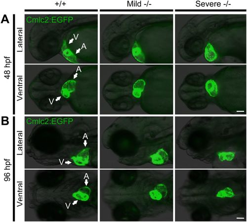
Pdgfra homozygous mutants exhibit abnormal cardiac phenotypes. An incross of the pdgfra gene-trapped strain carrying a Tg(cmlc2:EGFP) transgene was used to investigate the cardiac phenotype. (A) At 48?hpf, mild and severe ?/? mutants present with abnormal cardiac looping; severe ?/? embryos also develop an enlarged atrium and pericardial edema. (B) At 96?hpf, the hearts of mild ?/? mutants remain incompletely looped, while severe ?/? mutants present with collapsed chambers and pericardial edema. Arrows pointing at ventricle (V) and atrium (A). The fish cranium is on the left. Scale bar: 100??m. |
|
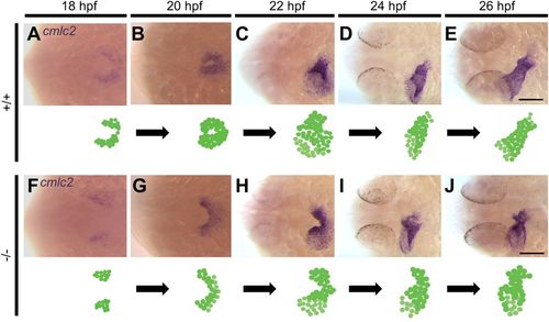
Homozygous pdgfra mutants demonstrate abnormal cardiac fusion. Representative photomicrographs of whole-mount in situ hybridization using the cardiac myosin light chain 2 (cmlc2) riboprobe to label myocardial cells. In +/+ embryos, the posterior regions of the bilateral cardiac fields interact during cardiac fusion at 18?hpf (A), followed by interaction between the anterior regions at 20?hpf (B). At 22?hpf, the cardiac tube forms and symmetry is broken by leftward displacement (jogging) (C). At 24 (D) and 26 (E) hpf, the cardiac tube elongates. In ?/? mutants, cardiac fusion is delayed at 18?hpf (F). Interaction between the posterior regions occurs at 20?hpf (G). At 22?hpf, the anterior regions of the bilateral cardiac fields fail to fuse (H). By 24 (I) and 26 (J) hpf, the anterior portions of the bilateral cardiac fields begin to come into contact. Dorsal views are shown, with the fish cranium on the left. An illustration of the developing heart is shown at the bottom of each figure. Scale bar: 100??m. EXPRESSION / LABELING:
PHENOTYPE:
|
|
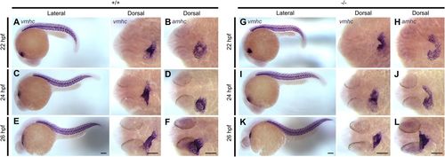
Delay in anterior cardiac fusion disrupts the development of cardiac chambers in pdgfra mutants. Representative images of embryos subjected to whole-mount in situ hybridization with riboprobes against ventricular myosin heavy chain (vmhc) and atrial myosin heavy chain (amhc). In +/+ embryos, the ventricular cells are observed at the apex of the cone (A) and the atrial cells at its base (B) at 22?hpf. At 24 and 26?hpf, the ventricles elongates (C and E), and the atria become cohesive and tubular (D and F). In ?/? mutants both the ventricular (G) and atrial (H) regions of the cardiac cone remain incompletely fused at 22?hpf. At 24 to 26?hpf, the anterior portions of the bilateral cardiac fields begin to come into contact; however, the ventricles (I and K) and the atria (J and L) appear shorter and wider. Lateral and dorsal views are shown, with the fish cranium on the left. Scale bar: 100??m. EXPRESSION / LABELING:
PHENOTYPE:
|
|
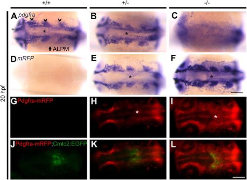
Pdgfra is expressed in the midline during cardiac assembly. Representative photomicrographs of whole-mount in situ hybridization using pdgfra and mRFP riboprobes at 20?hpf (A-F). In +/+ (A) and +/? (B) embryos expression of pdgfra is observed in different tissues, including the developing cranial ganglia (arrow head), anterior lateral plate mesoderm (ALPM) (arrow), optic cup (cross), and the midline (asterisk, also in panels E and F). No specific pdgfra expression is detected in ?/? embryos (C). Expression of mRFP is not detected in +/+ embryos (D). On the other hand, mRFP expression in +/? (E) and ?/? (F) embryos is similar to pdgfra expression. Representative images of 20?hpf live GBT1300 siblings carrying a Tg(cmlc2:EGFP) transgene (G-L). No Pdgfra-mRFP expression is observed in +/+ embryos (G). On the other hand, +/? (H) and ?/? (I) siblings show Pdgfra-mRFP expression similar to the mRNA expression data, including midline expression where cardiac assembly occurs (asterisk). Complete cardiac fusion is observed in +/+ (J) and +/? (K) embryos, while homozygous mutants demonstrated cardiac fusion only at the posterior end (L). Dorsal views are shown, with the fish cranium on the left. Scale bar: 75??m (A-F) and 250??m (G-L). EXPRESSION / LABELING:
PHENOTYPE:
|
|
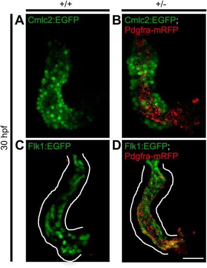
Analysis of the developing heart suggested expression of Pdgfra-mRFP in the endocardium rather than the myocardium. Dissected hearts from +/+ and +/? GBT1300 siblings carrying either a Tg(cmlc2:EGFP) or Tg(flk1:EGFP) transgene were used to investigate the expression pattern of the fusion Pdgfra-mRFP at 30?hpf. Cmlc2 positive +/+ embryos show no Pdgfra-mRFP expression in the heart (A), while +/? embryos exhibit Pdgfra-mRFP expression internal to the myocardium (B). Flk positive +/+ embryos show no Pdgfra-mRFP expression in the heart (C), while +/? embryos suggested co-localization of Pdgfra-mRFP with the endocardium (D). Scale bar: 50??m. EXPRESSION / LABELING:
|
|
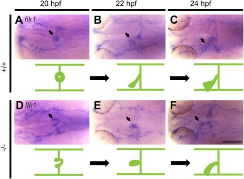
Migration of endocardial precursors to the midline is abnormal in pdgfra mutants. Representative photomicrographs of whole-mount in situ hybridization using fetal liver kinase 1 (flk1) riboprobe. In +/+ embryos, endocardial precursors migrate and fuse at the midline forming a ring-like structure at 20?hpf (A), followed by elongation and leftward movement at 22 (B) and 24 (C) hpf. In ?/? mutants, endocardial precursors form a V-like structure at 20?hpf (D). Endocardial cells reach the midline and begin to elongate at 22?hpf (E). Elongation continues with abnormal leftward movement at 24?hpf (F). Arrows point to endocardium. Dorsal views are shown, with the fish cranium on the left. An illustration of the developing heart is shown at the bottom of each figure. Scale bar: 100??m. EXPRESSION / LABELING:
PHENOTYPE:
|
|
Neural crest cells fail to reach the oral ectoderm in mild and severe pdgfra mutants. A cross of GBT1300 with a Tg(sox10:EGFP) transgenic line was used to investigate neural crest migration at 24 hpf. In contrast to +/+ embryos, Mild and severe -/- mutants demonstrate failure of neural crest cells to reach the oral ectoderm (arrow). The fish cranium is on the left. Scale bar: 100 ?m. |
|
Mild and severe pdgfra homozygous mutants present abnormal cardiac morphology. A cross of GBT1300 with a Tg(cmlc2:EGFP) line was used to investigate the cardiac phenotype at 96 hpf. In contrast to their +/+ siblings, mild and severe -/- mutants present abnormal cardiac looping and morphology. Ventral views are shown. The fish cranium is on the left. |
|
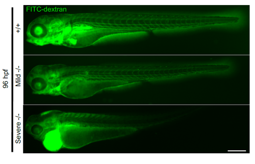
Severe homozygous pdgfra mutants lack blood circulation. Injection of FITC-dextran (4 KD) into the common cardinal vein reveals absence of circulation in severe -/- larvae, compared to +/+ and mild -/- larvae at 96 hpf. Lateral views are shown, with the fish cranium on the left. Scale bar: 250 ?m. PHENOTYPE:
|
|
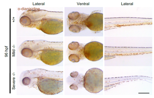
O-Dianisidine staining demonstrates presence of hemoglobinized blood in severe homozygous pdgfra larvae. Representative bright field images of OD-stained larvae indicating the presence of hemoglobinized blood in +/+, mild -/- and severe -/- larvae at 96 hpf. Lateral and ventral views are shown, with the fish cranium on the left. Scale bar: 250 ?m. |


