- Title
-
Endocytic Adaptor Protein Tollip Inhibits Canonical Wnt Signaling
- Authors
- Toru?, A., Szyma?ska, E., Castanon, I., Woli?ska-Nizio?, L., Bartosik, A., Jastrz?bski, K., Mi?tkowska, M., González-Gaitán, M., Miaczynska, M.
- Source
- Full text @ PLoS One

ZFIN is incorporating published figure images and captions as part of an ongoing project. Figures from some publications have not yet been curated, or are not available for display because of copyright restrictions. EXPRESSION / LABELING:
|
|
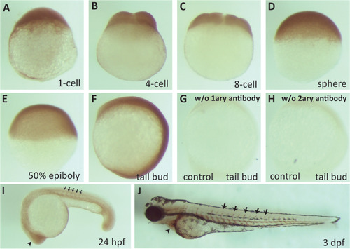
zTollip is broadly expressed during early zebrafish development. Immunocytochemistry analysis of zTollip protein localization in early embryonic development of zebrafish. Embryos fixed at various stages (indicated below the images) were incubated with primary antibodies against human Tollip, followed by secondary antibodies and chromogenic peroxidase-based detection. As controls for staining specificity, embryos at tail bud stage were processed omitting the primary antibody (G) or secondary antibody (H). zTollip expression is uniform from 1-cell stage until tail bud (A-F), but its expression increases in the head (arrowhead in I) and intersomitic regions (black arrows in I) at 24 hpf. At 3 dpf, zTollip is maintained in the intersomitic regions (arrows in J), but it is also expressed in the tissue surrounding the heart (arrowhead in J). Lateral views (A-H). Anterior to the left (I, J). EXPRESSION / LABELING:
|
|
zTollip contributes to the canonical Wnt pathway in embryonic development. (A-E) Embryos overexpressing ztollip (B) or injected with either ztollip morpholinos (MOs) (C), or β-catenin2 MO (D), or both (E) were analyzed for their morphological phenotype at 48 hpf. Typical phenotypes are shown. Lateral views with head to the left. Injection of ztollip mRNA led to reduction of posterior structures (arrow in B) compared with wild-type, control embryos (A). Knockdown of ztollip resulted in tail curvature (arrow in C) and pericardial oedema (asterisk in representative embryo in inset of C). Injection of β-catenin2 MO caused morphological defects similar to those found in embryos overexpressing ztollip (compare B and D). Tail curvature and pericardial oedema in embryos overexpressing ztollip could be rescued by injection of 2 ng of β-catenin2 MO (compare B and E). (F-R) In situ hybridization using different mRNA probes. Embryos injected with ztollip mRNA showed expanded expression of both the dorsal mesendodermal marker goosecoid (gsc) at 6 hpf (shield stage) (brackets in G) and the dorsal marker notail (ntl) at tail bud stage (brackets in I) compared to uninjected embryos (F and H, respectively). Expression of the cardiac marker cmlc2 (J, K) indicated the failure of the heart to form the loop in ztollip morphants (asterisk in J) compared to wild-type embryos (asterisk in K) at 48 hpf. Expression of fgf8 in the tail was reduced or absent in embryos overexpressing ztollip (arrow in M), while embryos injected with ztollip MOs showed an increase in fgf8 expression in the brain and in the tail (arrows in O) compared to uninjected, control embryos (L) at 24 hpf. Embryos injected with 8 ng β-catenin2 MO exhibited reduced fgf8 expression (N). Blocking endocytosis by injecting embryos with 4 ng rab5a MO or by expressing the Dnm2-K44A displayed no clear effect on fgf8 expression (arrows in P and Q). ztollip overexpression together with the Dnm2-K44A mutant showed a Tollip-like phenotype on the expression of fgf8 (arrow in R). |
|
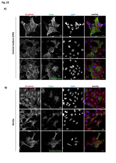
Intracellular distribution of β-catenin upon overexpression of Tollip. HEK293 cells transfected for 24 h with myc-tagged Tollip: wild-type (wt), del.1 deletion mutant or M240A/F241A point mutant were stimulated with control medium (CM; A) or Wnt3a-conditioned medium (B) for 8 h. Cells were immunostained for β-catenin and myc tag (overexpressed Tollip), with DAPI indicating cell nuclei. Scale bar 20 µm. |
|
Dose responses to the β-catenin2 morpholino. Zebrafish embryos were injected at the 1-cell stage with β-catenin2 morpholino (MO) at different concentrations (2 ng, 4 ng, 8 ng, 16 ng). Phenotypes of injected embryos at 48 hpf are shown, compared to an uninjected wild-type embryo. Lateral views with head to the left. |


